IMAGE

Fig. 4

ID
ZDB-IMAGE-210619-9
Source
Figures for Zou et al., 2021
Image
Figure Caption

Fig. 4

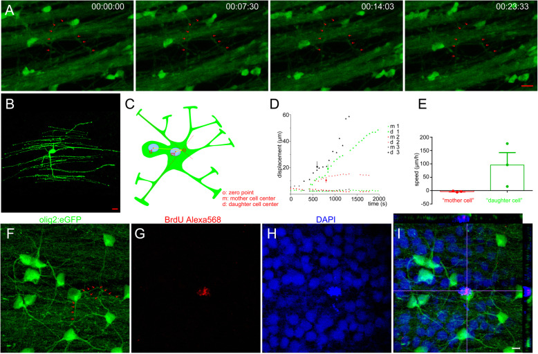
mOLs within the retina are divided in an asymmetric manner. a Serial imaging showed that the mOL within the retina was under division. Arrows indicate the processes that maintained integrity during the whole imaging. b The olig2+ cell within the retina is in a mature state, as shown by the perfect myelin structure. c Schematic picture shows the method to calculate cell displacement in the asymmetric division of mOL. The origin point is the crossover of two random myelin processes. d The displacement of the mother and daughter cells in the asymmetric division of mOLs within the retina. Arrows show the time point at which division was completed. e Speeds of mother and daughter cells. Daughter cells had a high migration ability while mother cells did not (p < 0.05). f-i BrdU results show that a mOL within the retina was proliferating, while the myelin structure remained integral (arrows). The image has 10 slices, and the interval is 0.5 μm. Scale bar: 10 μm (a-b); 5 μm (f-i)

Acknowledgments
This image is the copyrighted work of the attributed author or publisher, and ZFIN has permission only to display this image to its users. Additional permissions should be obtained from the applicable author or publisher of the image. Full text @ Cell Regen (Lond)