IMAGE

Fig. 2

ID
ZDB-IMAGE-201216-5
Source
Figures for Vallese et al., 2020
Image
Figure Caption

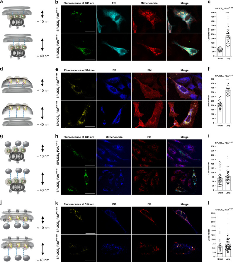
Fig. 2 Functional characterization of the SPLICS<sub>S/L</sub>-P2A probes.

a, d, g, j Cartoon showing the short- and long-range contact sites measured by the indicated reporters. b, e, h, k Representative images of HeLa cells expressing the indicated SPLICSS/L-P2A probes to identify the co-localization of GFP (488 nm) or YFP (514 nm) fluorescent dots with targeted organelles. Different markers are used to label organelles: red fluorescent protein (mCherry) targeted to PM, anti-KDEL antibody (ER), mtHSP60 (mitochondria) and anti-PMP70 antibody (PO). Confocal images were acquired at 405, 488, 514, and 588 nm excitation wavelength. Scale bar 25 µm. c, f, i, l Quantification of the indicated SPLICSS/L-P2A (short and long) contacts in HeLa cells. The SPLICS dots were quantified from the 3D rendering of a complete Z-stack (see Supplementary Fig. 2), mean ± SEM. The data were obtained from three independent transfections. (****P ≤ 0.0001 unpaired two-tailed t-test). Source data are provided as a source data file.

Acknowledgments
This image is the copyrighted work of the attributed author or publisher, and ZFIN has permission only to display this image to its users. Additional permissions should be obtained from the applicable author or publisher of the image. Full text @ Nat. Commun.