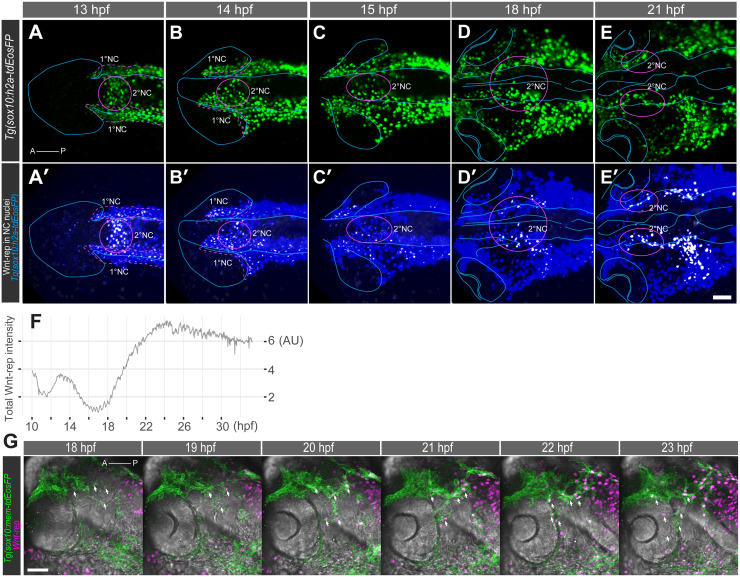
Dorsal views of a Tg(sox10:h2a-tdEosFP;wnt-rep) embryo highlighting (A-E) h2a-tdEosFP-positive NC cells and (A’-E’) Wnt reporter expression (white) in h2a-tdEosFP-positive NC cells (blue) showing colocalized Wnt reporter with nuclei from sox10-positive NC cells. At 13–14 hpf, 2°NC cells in the medial region (magenta circles, A-B) express Wnt-rep (A’-B’) at higher levels than 1°NC cells. This Wnt-rep expression is transient and followed by a reduction at 15 hpf (C’). At 18–21 hpf, 2°NC cells develop into bilateral clusters at the level of the diencephalon and mesencephalon (magenta circles, D-E). These cells express high levels of Wnt-rep (D-E’). Orientation: anterior left. (F) Mean total intensity (expressed in arbitrary unit, AU) profile of Wnt reporter expression in sox10-NC cells during 10–33 hpf shows 1st and 2nd wave of Wnt activity. (G) 2°NC cells (green) express Wnt reporter (magenta) prior to and during migration into the distal side of the eye (arrows). Orientation: anterior left, dorsal up; Scale bars: 50 μm.
Acknowledgments
This image is the copyrighted work of the attributed author or publisher, and
ZFIN has permission only to display this image to its users.
Additional permissions should be obtained from the applicable author or publisher of the image.
Full text @ PLoS Genet.
Your Input Welcome
Thank you for submitting comments. Your input has been emailed to ZFIN curators who may contact you if
additional information is required.
Oops. Something went wrong. Please try again later.