
Fig. 4
- ID
- ZDB-IMAGE-200424-43
- Genes
- Publication
- Guglielmi et al., 2020 - Temporal control of Wnt signaling is required for habenular neuron diversity and brain asymmetry
- All Figures
- Figures for Guglielmi et al., 2020
|
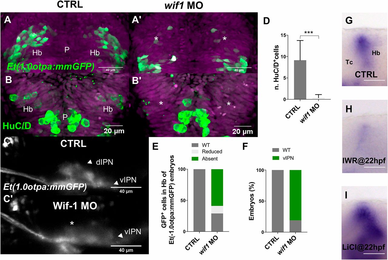
Fig. 4 Wif1 controls dHb neuron differentiation and is regulated by Wnt signaling. (A-C′) Projections of confocal z-stacks. (A-B′) Dorsal views, anterior is towards the top focused onto the diencephalon of embryos at 3 dpf. Nuclei are DAPI labeled (purple). (A-B′,D,E) Wif1 hypomorphic embryos exhibit delayed habenular neuron differentiation (asterisks in A′,B′). (C,C′) Lateral views focused on the IPN of Et(-1.0otpa:mmGFP) embryos with anterior towards the left. (C,C′,F) Wif1 knockdown embryos show predominant vIPN innervation. Asterisk highlights the missing dIPN innervation. (G-I) Lateral views focused on the diencephalon of 25 hpf (G) untreated and (H,I) treated embryos, labeled for Wif1 expression. Scale bar: 60 µm. Labeling procedures were conducted in parallel for comparison. d, dorsal; Hb, habenula, IPN, interpeduncular nucleus; P, pineal; v, ventral.