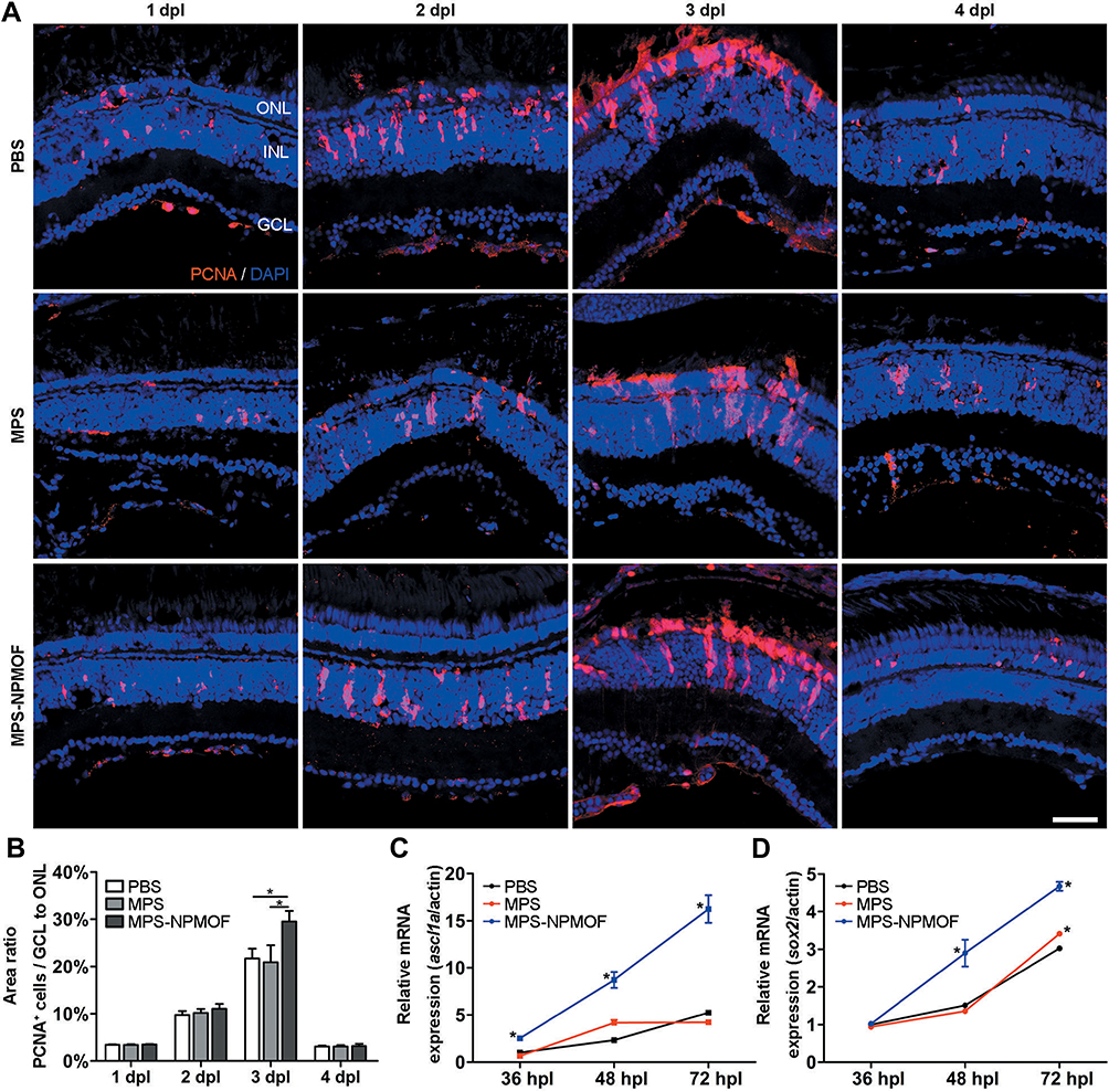
Fig. 7 Cell proliferation of light-lesioned retina following injection of MPS-NPMOF. (A) PCNA staining in sections taken from retinas at 1, 2, 3 and 4 dpl. (B) Statistical analysis of the percentage of PCNA-positive cells from GCL to ONL. There was a significantly higher percentage in the MPS-NPMOF group than in the PBS and MPS groups at 3 dpl (ANOVA, *p<0.05). (C and D) The expression of ascl1a and sox2 mRNA. Note that ascl1a was increased in the MPS-NPMOF group at 36, 48 and 72 hpl, while sox2 was increased at 48 and 72 hpl (ANOVA, *p<0.05). Scale bar in (A): 20 μm. Abbreviations: ONL, outer nuclear layer; INL, inner nuclear layer; GCL, ganglion cell layer.
Image
Figure Caption
Acknowledgments
This image is the copyrighted work of the attributed author or publisher, and
ZFIN has permission only to display this image to its users.
Additional permissions should be obtained from the applicable author or publisher of the image.
Full text @ Int. J. Nanomedicine

