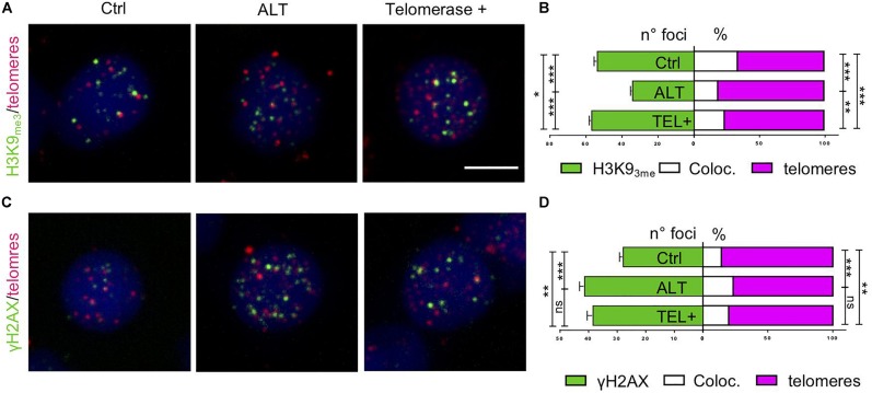
Expression of tert maintains telomeric heterochromatin in brain tumors. (A,C) Fluorescent microscope images of representative control, ALT, and telomerase + zebrafish brain tumor cells, stained via immunofluorescence (green) combined with telomere-FISH (magenta). Antibody against chromatin methylation marks (A, H3K9me3) and DNA damage marker (C, γH2AX), were used and counterstained with DAPI. Scale bar: 5 μm. (B,D) Immunofluorescence quantification expressed as the number of foci per nucleus (green), and percent of immunofluorescence foci (white) that colocalized at telomeres (magenta) per nucleus (n = 25–60 nuclei) for the corresponding images in panel (A,C). *p < 0.05, ***p < 0.001 between the indicated groups. Bars represent mean ± SEM.
Acknowledgments
This image is the copyrighted work of the attributed author or publisher, and
ZFIN has permission only to display this image to its users.
Additional permissions should be obtained from the applicable author or publisher of the image.
Full text @ Front Cell Dev Biol
Your Input Welcome
Thank you for submitting comments. Your input has been emailed to ZFIN curators who may contact you if
additional information is required.
Oops. Something went wrong. Please try again later.