IMAGE

Figure 5

ID
ZDB-IMAGE-200302-50
Source
Figures for Otero-Asman et al., 2020
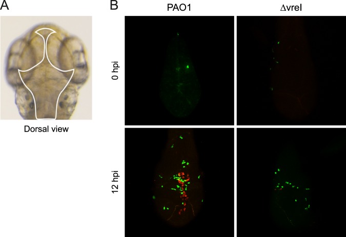
Image
Figure Caption

Figure 5

σVreI activation during P. aeruginosa infection in zebrafish embryos. (A) Dorsal view of the head of a two days post-fertilization zebrafish embryo. The hindbrain ventricle where P. aeruginosa was injected is highlighted. (B) Confocal images of the head of embryos injected in the hindbrain ventricle with ∼2000 CFU of the P. aeruginosa PAO1 wild-type strain or its isogenic ΔvreI mutant bearing the σVreI-dependent pdtA::rfp transcriptional fusion (pMP0690mCherry plasmid, Table S1) (red channel) at 0 and 12 hpi. P. aeruginosa was grown in high Pi medium prior injection. Neutrophils expressing constitutively a green fluorescence protein (gfp) are also visualized (green channel). Images are representative of three independent experiments (N = 3).

Figure Data
Acknowledgments
This image is the copyrighted work of the attributed author or publisher, and ZFIN has permission only to display this image to its users. Additional permissions should be obtained from the applicable author or publisher of the image. Full text @ Sci. Rep.