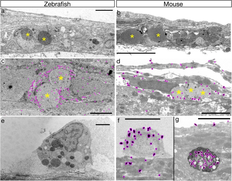
Ultrastructural properties of BLECs/LLECs are conserved in zebrafish and mice. a Representative TEM micrograph of adult zebrafish meninges. The large, round, densely and homogenously-stained inclusions (yellow asterisks) of a putative BLEC are indicated. Scale bar = 2.5 µm (in all images). n = 2 brains. b Representative TEM micrograph of adult mouse leptomeninges. Putative LLECs have their large inclusions marked with yellow asterisks. n = 2 brains. c Cells with large inclusions (yellow asterisks) characteristic of BLECs are immunologically labelled with post-embed detection of GFP in Tg(flt4:gfp) adult brains using a 20 nm gold-labelled secondary. Minimal contrast staining was used to prevent obscuring of the nanogold particles (highlighted in magenta). n = 2 brains. d Mouse meningeal cells with large inclusions (yellow asterisks) are labelled by pre-embed detection of LYVE1 using a nanogold secondary with silver-enhancement. The silver grains are highlighted with magenta. Note the LYVE1-positive cell has silver grains present near the outer membrane. n = 2 brains. e An example of a GFP-negative cell in zebrafish Tg(flt4:gfp) adult meninges with similar ultrastructural properties to macrophages. n = 2 brains. f A second population of LYVE1-positive cells in the mouse leptomeninges contain multiple small, irregular, heterogeneous vesicles with silver grains (magenta) present within the cytoplasm and vesicles. These cells are ultrastructurally similar to macrophages (e). n = 2 brains
Acknowledgments
This image is the copyrighted work of the attributed author or publisher, and
ZFIN has permission only to display this image to its users.
Additional permissions should be obtained from the applicable author or publisher of the image.
Full text @ Acta Neuropathol.
Your Input Welcome
Thank you for submitting comments. Your input has been emailed to ZFIN curators who may contact you if
additional information is required.
Oops. Something went wrong. Please try again later.