
Fig. S6
- ID
- ZDB-IMAGE-190715-10
- Genes
- Publication
- Williams et al., 2018 - Requirement of zebrafish pcdh10a and pcdh10b in melanocyte precursor migration
- All Figures
- Figures for Williams et al., 2018
|
Fig. S6
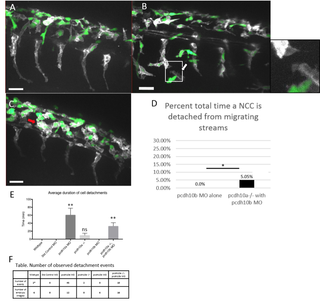
Knockdown of pcdh10b in pcdh10a mutants restores NCC migration defect. Lateral views of double transgenic zebrafish Tg(sox10:mRFP,mitfa::GFP), membrane RFP has been changed to grayscale. (A) Control wildtype embryos injected with pcdh10b MO NCCs 28hpf. (B, C) Lateral view of pcdh10a mutants injected with pcdh10b MO NCCs at 28hpf show defects in NCC migration, including in mitfa+ melanoblasts. White arrows mark NCCs that have detached from other NCCs in the migrating streams. Red arrows mark NCCs that have aggregated within the dorsal domain. (D) Percent of total time NCCs are detached from other NCCs in pcdh10b MO alone and pcdh10a-/- injected with pcdh10b MO embryos. * denotes P value of .035. Scale bar is 30um. (E) Quantification of the duration of cell detachment events across all samples, ** denotes P value of .006 (F) Table, comparison of the number of embryos imaged and the number of detachment events that occurred across all samples.
Reprinted from Developmental Biology, 444 Suppl 1, Williams, J.S., Hsu, J.Y., Rossi, C.C., Artinger, K.B., Requirement of zebrafish pcdh10a and pcdh10b in melanocyte precursor migration, S274-S286, Copyright (2018) with permission from Elsevier. Full text @ Dev. Biol.