
Fig. 1
- ID
- ZDB-IMAGE-190423-1
- Genes
- Publication
- Li et al., 2018 - Endodermal pouch-expressed dmrt2b is important for pharyngeal cartilage formation.
- All Figures
- Figures for Li et al., 2018
|
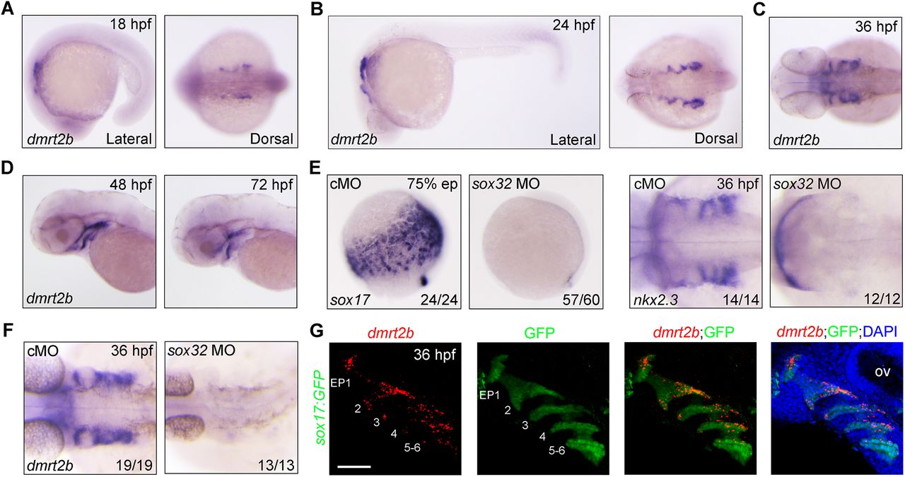
Fig. 1 Expression of dmrt2b in the developing endodermal pouches. (A–D) Analysis of dmrt2b expression at different stages. (E,F) Endodermal cells were absent from sox32 morphants. Expression of endodermal marker sox17 (E), endodermal pouch marker nkx2.3 (E) and dmrt2b (F) were examined by in situ hybridizations at the indicated stages in wild-type embryos injected with 8 ng control MO (cMO) or sox32 MO. (G) Expression of dmrt2b in endodermal pouches. At 36 hpf, Tg(sox17:GFP) transgenic embryos were stained for dmrt2b mRNA with Dr-dmrt2b-C3 probe (red), and then immunostained with anti-GFP antibody (green). Nuclei were counterstained with DAPI (blue). The six endodermal pouches are labeled in the left two panels. EP, endodermal pouch; ov, otic vesicle. Scale bar: 50 µm.