
Fig. 5
|
Fig. 5
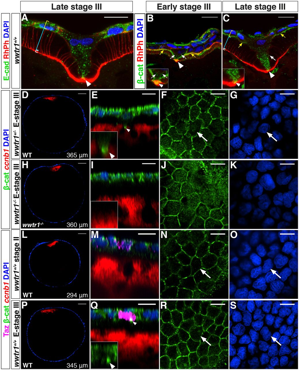
Taz is enriched in the MC precursor before detectable morphological changes. (A-C) Single plane confocal images of cryosectioned ovaries showing E-cadherin (A, n=8) or β-catenin (B,C, n=10) localization in the MC and the neighboring FCs at the contact point (white arrowhead), at the MC membranes (white arrows) and at the interface between the oocyte microvilli and the FCs (yellow arrows). White, cyan and yellow brackets indicate the oocyte cortex, the microvilli/vitelline membrane and the FCL, respectively. Insets (B,C) are higher magnification views of the contact point. Rhodamine Phalloidin (RhPh) and DAPI were used to independently mark membranes and nuclei, respectively. (D-S) Confocal images of wwtr1+/+, wwtr1+/− or wwtr1−/− whole-mount follicles at the indicated stage stained with Taz and/or β-catenin antibodies, ccnb1 mRNA and DAPI as indicated. D,H,L,P are overview images with the follicle size indicated in the lower right corner. White arrowheads in E and Q point to β-catenin enrichment at the contact point; white arrows indicate an MC. E-stage III, early stage III. Scale bars: 10 μm in A-C,E-G,I-K,M-O,Q-S; 50 μm in D,H,L,P.