
|
Fig. S1
Role of Shh signaling in the waves of adaxial cell elongation along D-V axis, related to Figure 2
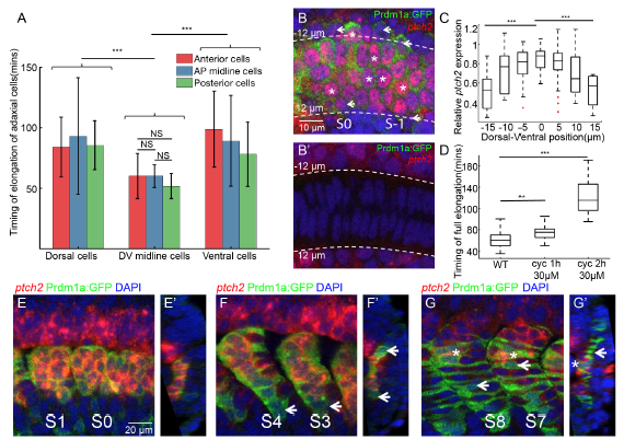
(A) The timing of elongation of adaxial cells at different AP and DV positions. Red, blue and green denote adaxial cells from anterior-most part, central part and posterior-most part of adaxial cell population ( nCells=135 from total of 8 somites from 8 different embryos). (B-B’) Fluorescent in situ of ptch2 at PSM on the plane of adaxial cells (B) or the notochord (B’). White short arrow and white asterisks denote the adaxial cells with relative low and high ptch2 expression respectively. Images are taken at 18-somite stage at PSM. (C) Ptch2 expression level per cell along the DV axis. (nCells=101 from total of 5 somites from 5 different embryos). Adaxial cells are labeled with Prdm1a:GFP. (D) Timing of elongation of midline adaxial cells in wild type embryos (nCells=37), embryos under 1 hour (nCells=34) or 2 hours (nCells=47) cyclopamine treatment at 30μM. The cyclopamine treatment was performed at 1 hour or 2 hours before the start of live-imaging. Adaxial cells of somite S0 were then quantified for their timing of elongation. (E-G) Fluorescent in situ of ptch2 within adaxial cells and slow muscle fibers at different stages: (E) PSM and S1; (F) S3 and S4; (G)S7 and S8. (E’-G’) Constructed transverse images of (E-G) with dorsal to the top and medial to the left. White arrows and asterisks denote SSFs that had losing ptch2 expression and MPs that sustained ptch2 expression respectively. Adaxial cells and Slow muscles are labeled with Prdm1a:GFP. ***p < 0.001, NSP >0.05, Student’s t test.
Reprinted from Developmental Cell, 46, Yin, J., Lee, R., Ono, Y., Ingham, P.W., Saunders, T.E., Spatiotemporal Coordination of FGF and Shh Signaling Underlies the Specification of Myoblasts in the Zebrafish Embryo, 735-750.e4, Copyright (2018) with permission from Elsevier. Full text @ Dev. Cell