
Fig. 5
- ID
- ZDB-IMAGE-181019-35
- Publication
- Yin et al., 2018 - Spatiotemporal Coordination of FGF and Shh Signaling Underlies the Specification of Myoblasts in the Zebrafish Embryo
- All Figures
- Figures for Yin et al., 2018
|
Fig. 5
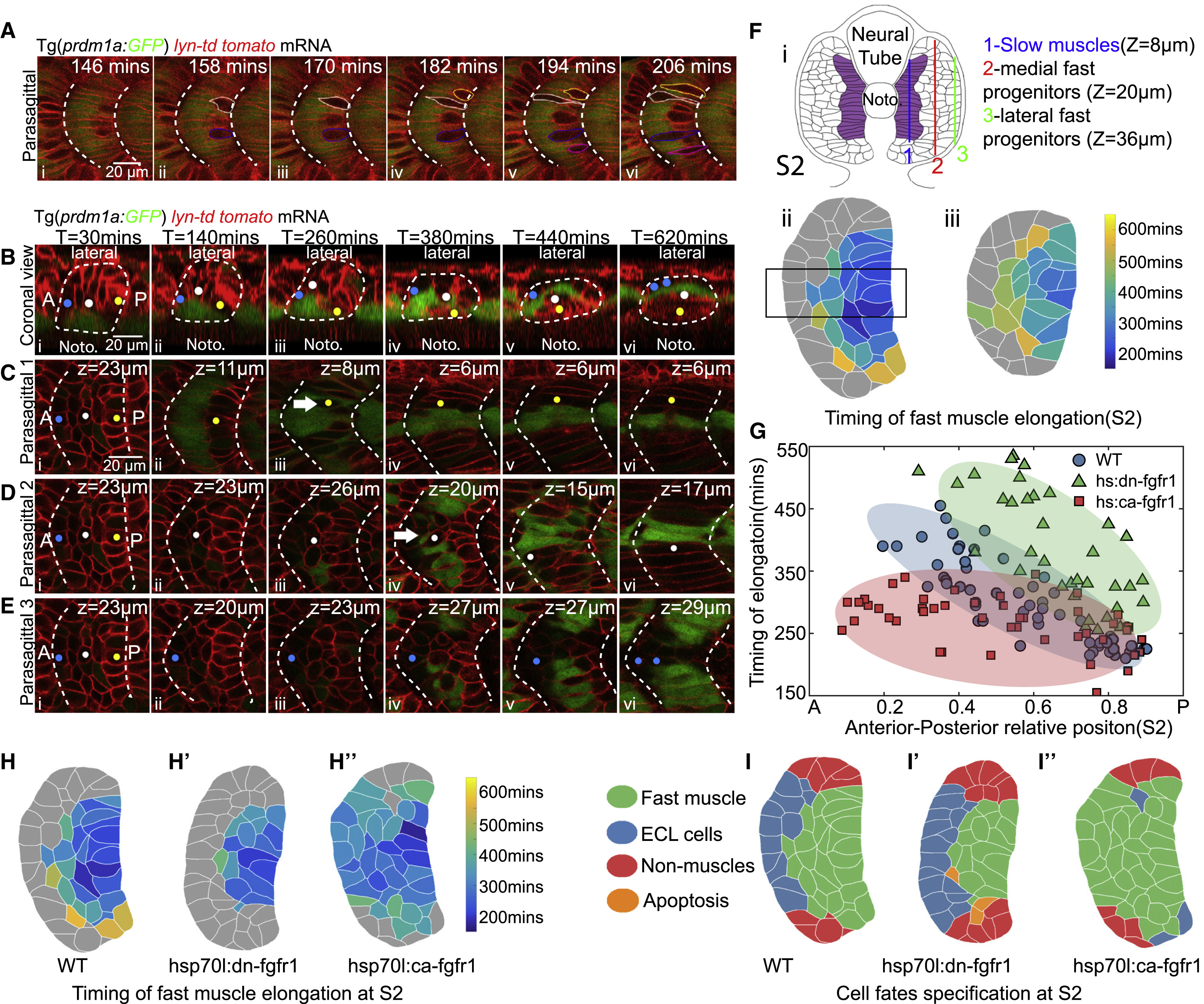
AP Polarity of Fast Muscle Myogenesis and the Roles of FGF Signaling in the AP Patterning
(A) Time-lapse of the earliest fast muscle elongation and the displacement of slow muscles. Yellow, white, blue, and magenta labels the contours of elongating fast muscle progenitors. Slow muscles are labeled with Prdm1a:GFP.
(B–E) Time-lapse of the cell rearrangement and differentiation of lateral somitic cells throughout the primary myogenesis at coronal view (B) with lateral to the top and anterior to the left and parasagittal optical views (C–E). Yellow, white, and blue circles label cells from posterior (C), central (D) and anterior (E) part of somite S2. In (C)–(E), the z-plane selected corresponds to the position of the center of each cell being tracked; corresponding z-position is clear in (B). White arrows label the elongating fast muscle progenitors.
(F) Maps of the timing of fast muscle elongation constructed at somite stage S2 (i) at parasagittal planes 20 μm (ii) or 36 μm (iii) away from the notochord. Cells labeled with gray color did not elongate throughout the primary myogenesis.
(G) The timing of elongation of fast muscle progenitors quantified from the rectangle region of (Fii) along the AP axis in wild-type embryo (blue, nCells = 57 from total of 7 somites taken from 6 embryos), embryos under heat shock of hsp70l:dn-fgfr1-eGFP (green, nCells = 35 from total of 6 somites taken from 5 embryos), or heat shock of hsp70l:ca-fgfr1 (red, nCells = 45 from total of 6 somites taken from 5 embryos). The ellipse represents the minimum-volume covering of data points in each group.
(H and I) Maps of timing of fast muscle elongation (H–Hʹʹ) and cell fates (I–Iʹʹ) at somite stage S2 in in wild-type embryo (H and I), embryos under heat shock of hsp70l:dn-fgfr1-eGFP (Hʹ and Iʹ), or heat shock of hsp70l:ca-fgfr1 (Hʹʹ and Iʹʹ). Images above are taken at somite 16–18.
Reprinted from Developmental Cell, 46, Yin, J., Lee, R., Ono, Y., Ingham, P.W., Saunders, T.E., Spatiotemporal Coordination of FGF and Shh Signaling Underlies the Specification of Myoblasts in the Zebrafish Embryo, 735-750.e4, Copyright (2018) with permission from Elsevier. Full text @ Dev. Cell