
|
Fig. 1
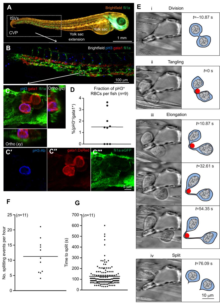
In vivo proliferation and splitting of circulating red blood cells (RBCs) in zebrafish embryos. (A) Overview of a zebrafish embryo at 48 hours post-fertilization (hpf) in vivo. ISVs, intersegmental vessels; CVP, caudal vein plexus. (B) Phospho-histone H3 (pH3) staining (blue) of the zebrafish caudal vein plexus. In Tg(fli1a:GFP;gata1:DsRed) double transgenic animals, endothelial cells (green) and RBCs (red) are labeled through expression of fluorescent markers as indicated. (C) Example of a pH3-positive circulating RBC. (D) Average extent of pH3-positive cells as percentage of circulating gata1-positive RBCs (n=9; >100 quantified RBCs per animal). (E) Splitting of proliferating RBCs is a four-step process. Following cell division (i), the two somata of a dividing RBC might eventually tangle (ii) at a vascular bifurcation which leads to the subsequent elongation (iii) of the soma-to-soma connection and its eventual rupture (iv). (F) We observed 11.24±0.93 RBC splitting events per hour (n=11; 142 events in total). (G) On average, a splitting event (i.e. phase ii–iv) occurred within 129.56±1.29 s (n=11; 142 events).