
IMAGE
Fig. S1
- ID
- ZDB-IMAGE-180912-17
- Genes
- Publication
- Kinoshita et al., 2018 - Functional roles of the Ripply-mediated suppression of segmentation gene expression at the anterior presomitic mesoderm in zebrafish
- All Figures
- Figures for Kinoshita et al., 2018
Image
|
Figure Caption
Fig. S1
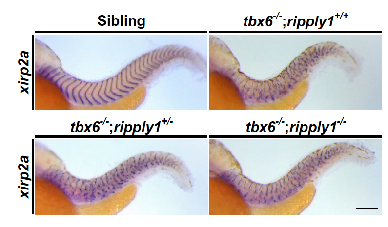
Defective somite segmentation is comparable between tbx6-/-; ripply1+/+and tbx6-/-; ripply1-/-embryos.
The segment boundaries oftbx6-/-; ripply1+/+, tbx6-/-; ripply1+/-, and tbx6-/-; ripply1-/-embryos at 36 hpfwere visualized by in situ staining using xirp2a/cb1045 probe. Lateral views and anterior is to the left. The photos were taken at the same magnification. Scale bar is 100 mm.
Figure Data
Acknowledgments
This image is the copyrighted work of the attributed author or publisher, and
ZFIN has permission only to display this image to its users.
Additional permissions should be obtained from the applicable author or publisher of the image.
Reprinted from Mechanisms of Development, 152, Kinoshita, H., Ohgane, N., Fujino, Y., Yabe, T., Ovara, H., Yokota, D., Izuka, A., Kage, D., Yamasu, K., Takada, S., Kawamura, A., Functional roles of the Ripply-mediated suppression of segmentation gene expression at the anterior presomitic mesoderm in zebrafish, 21-31, Copyright (2018) with permission from Elsevier. Full text @ Mech. Dev.