
Fig. 2
- ID
- ZDB-IMAGE-180912-12
- Genes
- Antibodies
- Publication
- Kinoshita et al., 2018 - Functional roles of the Ripply-mediated suppression of segmentation gene expression at the anterior presomitic mesoderm in zebrafish
- All Figures
- Figures for Kinoshita et al., 2018
|
Fig. 2
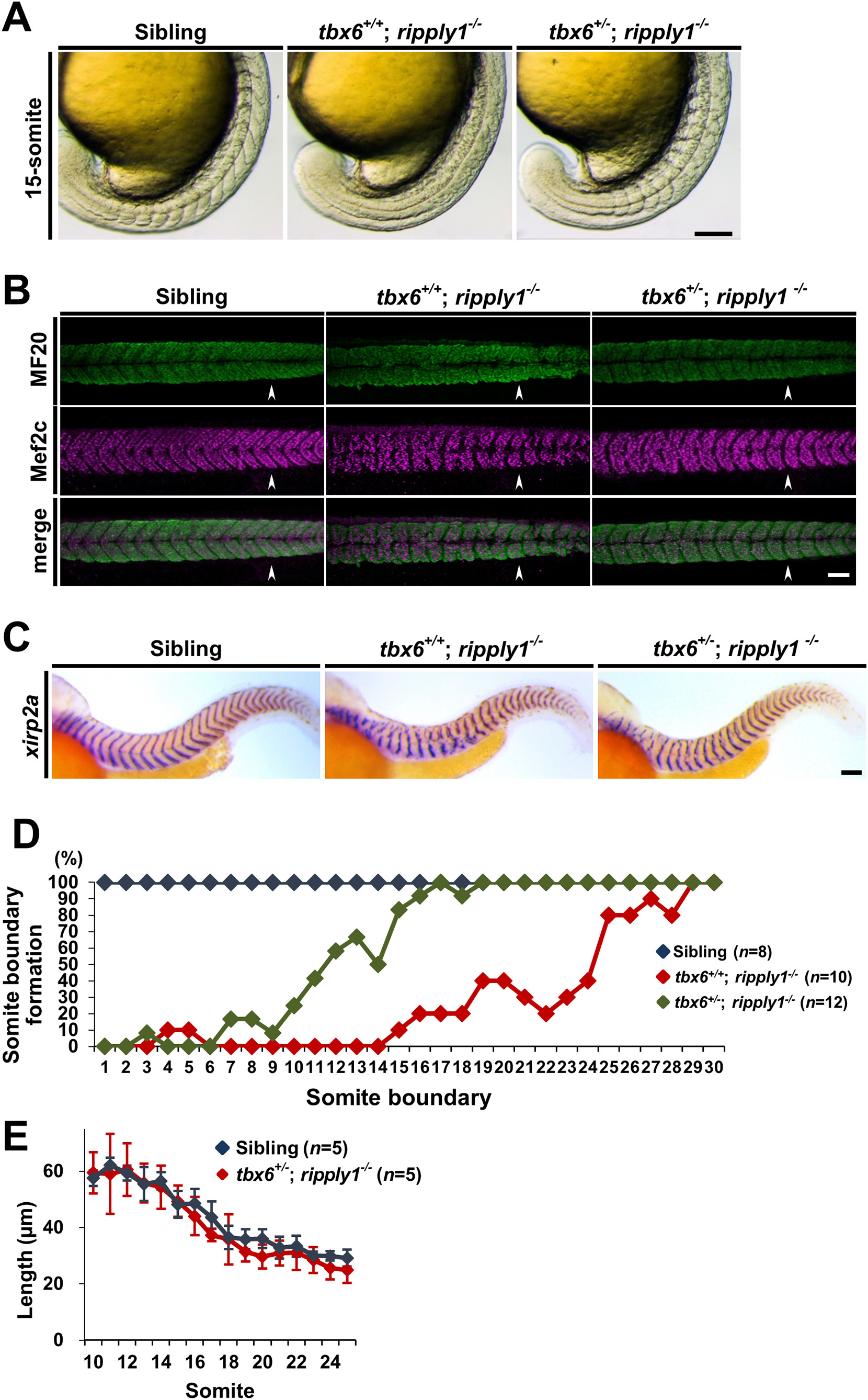
Heterozygous mutation of tbx6 in ripply1 homozygous mutants partially suppresses the defects of somite segmentation. (A) Lateral views of sibling, tbx6+/+; ripply1−/−, and tbx6+/−; ripply1−/− embryos at the 15-somite stage that were obtained by intercrosses between tbx6 and ripply1 double heterozygous mutants. After taking photos, the genotype of each embryo was determined by the heteroduplex mobility shift assay or DNA sequencing. (B) Immunostaining showing myotomes of sibling, tbx6+/+; ripply1−/−, and tbx6+/−; ripply1−/− embryos at 36 hpf using MF20 (myosin heavy chain, green) and anti-Mef2c (Myocyte enhancer factor 2, magenda) antibodies. Arrowhead indicates the posterior end of yolk extension. Anterior is to the left and dorsal is to the top. (C) Representative images of sibling, tbx6+/+; ripply1−/−, and tbx6+/−; ripply1−/− embryos at 36 hpf stained with a segment boundary marker, xirp2a/cb1045. Lateral views and anterior is to the left. (D) Distribution of somite boundary formation in sibling, tbx6+/+; ripply1−/−, and tbx6+/−; ripply1−/− embryos at 36 hpf. See Materials and methods. (E) The horizontal lengths of the somites were compared between sibling and tbx6+/−; ripply1−/− embryos. After staining with xirp2a, the intervals of somites (10th–25th segments) along the horizontal myoseptum of sibling and tbx6+/−; ripply1−/− embryos at 36 hpf were measured by ImageJ software. Scale bars in A, B, and C are 100 μm.
Reprinted from Mechanisms of Development, 152, Kinoshita, H., Ohgane, N., Fujino, Y., Yabe, T., Ovara, H., Yokota, D., Izuka, A., Kage, D., Yamasu, K., Takada, S., Kawamura, A., Functional roles of the Ripply-mediated suppression of segmentation gene expression at the anterior presomitic mesoderm in zebrafish, 21-31, Copyright (2018) with permission from Elsevier. Full text @ Mech. Dev.