
Fig. 3
- ID
- ZDB-IMAGE-180911-22
- Genes
- Publication
- Guner-Ataman et al., 2018 - Failed Progenitor Specification Underlies the Cardiopharyngeal Phenotypes in a Zebrafish Model of 22q11.2 Deletion Syndrome
- All Figures
- Figures for Guner-Ataman et al., 2018
|
Fig. 3
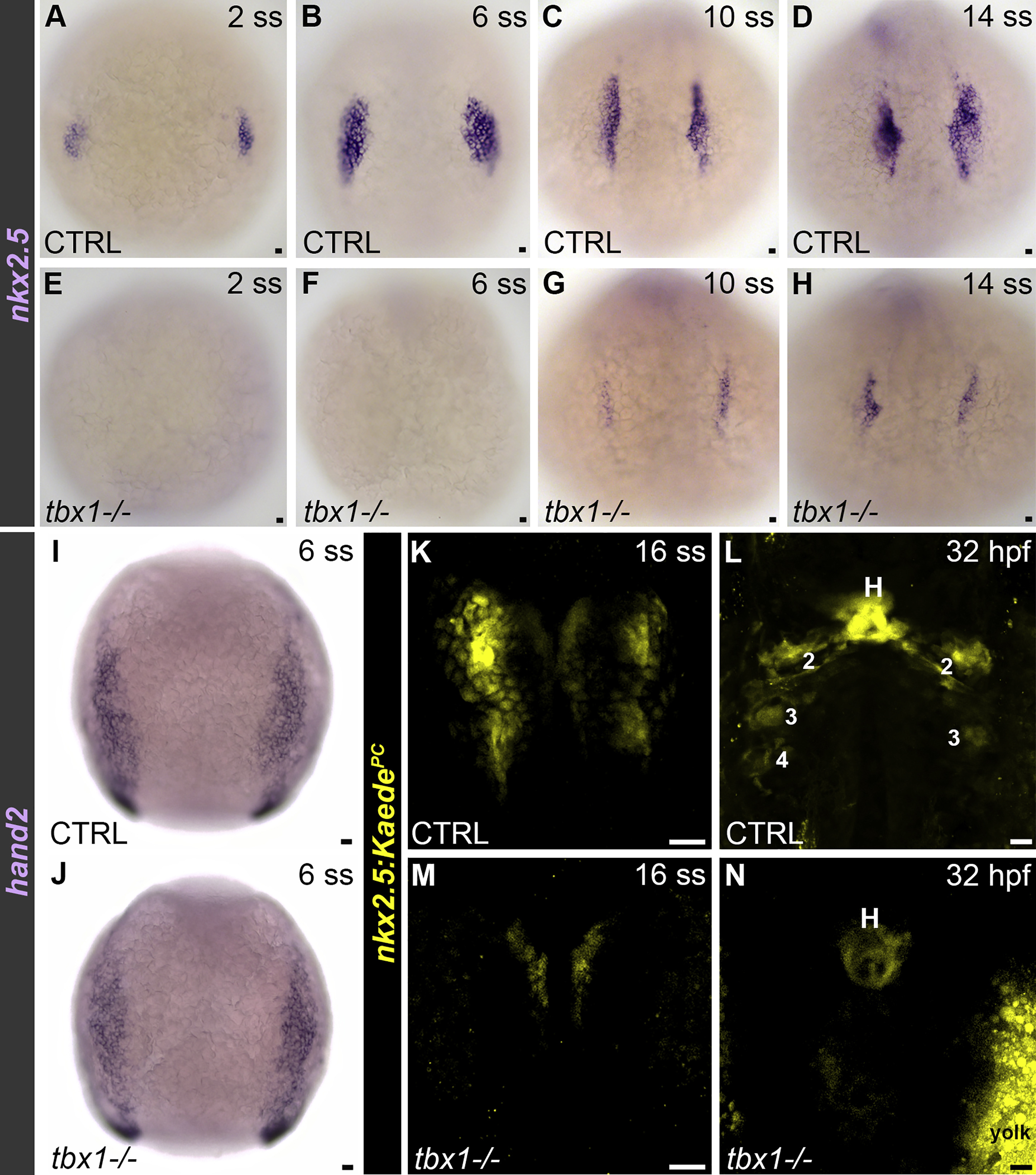
Tbx1 Is Required for Specification of the nkx2.5+ Pharyngeal Lineage in the ALPM of Zebrafish Embryos
(A–H) Bright-field images of 2 somite stage (ss) (A, n = 30; E, n = 10), 6 ss (B, n = 60; F, n = 20), 10 ss (C, n = 30; G, n = 11), and 14 ss (D, n = 10; H, n = 10) control (CTRL) (A–D) and tbx1 mutant (E–H) embryos processed by in situ hybridization with an nkx2.5 riboprobe. Dorsal views, anterior up.
(I and J) Bright-field z-stacks of 6 ss CTRL (I; n = 42) and tbx1 mutant (J; n = 13) embryos processed by in situ hybridization with a hand2 riboprobe. Dorsal views, anterior up.
(K–N) Confocal z-stacks of CTRL (K and L) and tbx1 mutant (M and N) Tg(nkx2.5:Kaede) embryos imaged immediately following photoconversion at the 16 ss (K and M) and again at 32 hr post-fertilization (L and N). Four of four CTRL embryos contained photoconverted Kaede protein (KaedePC, pseudocolored yellow) in the heart and pharyngeal clusters at 32 hpf. Four of four mutant embryos contained KaedePC exclusively in the heart. Dorsal views, anterior up.
In (A)–(K), little to no variation was observed between animals in each experimental group.
Scale bars, 25 μm.