
Fig. 2
- ID
- ZDB-IMAGE-180711-26
- Genes
- Publication
- Selland et al., 2018 - Coordinate regulation of retinoic acid synthesis by pbx genes and fibroblast growth factor signaling by hoxb1b is required for hindbrain patterning and development
- All Figures
- Figures for Selland et al., 2018
|
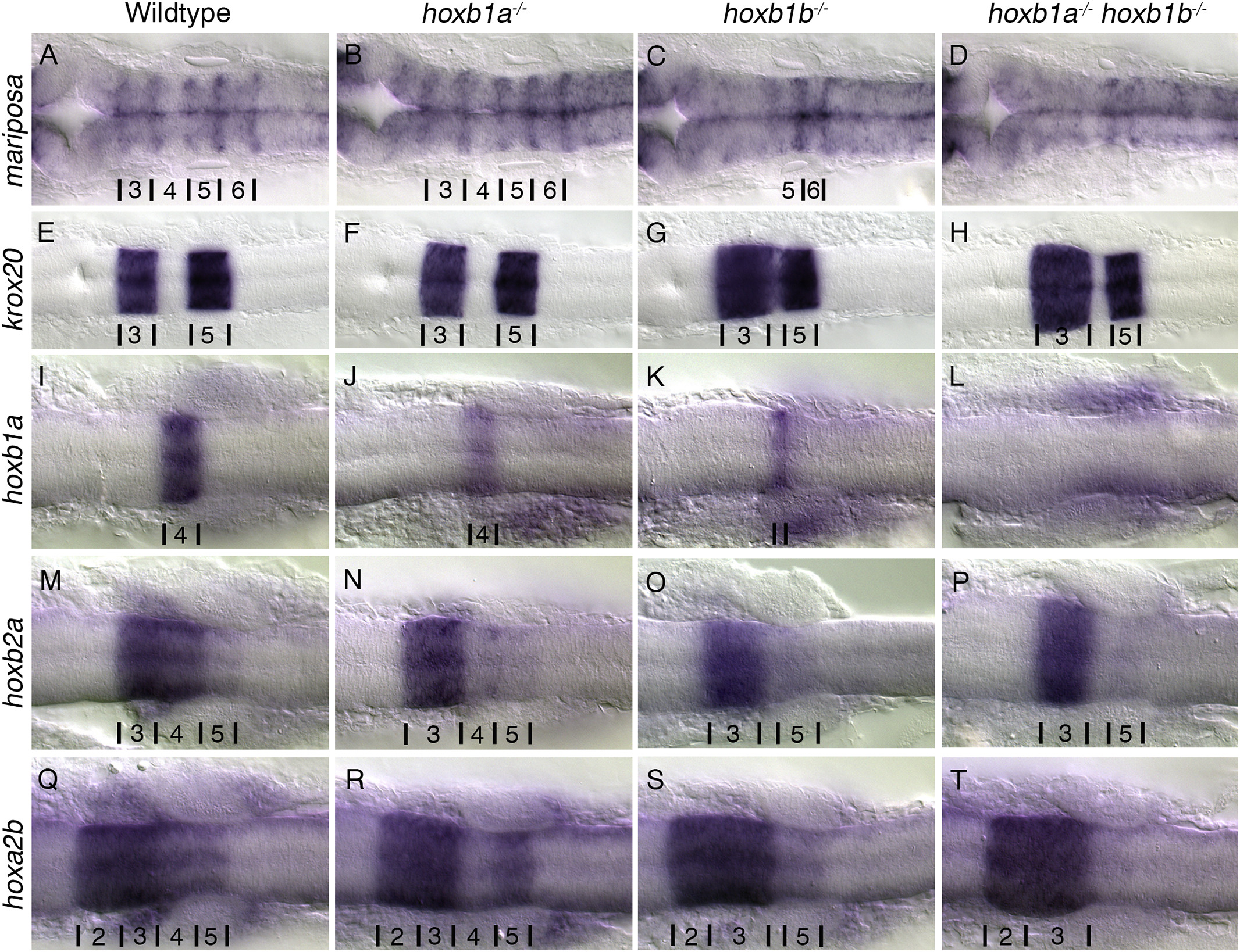
Fig. 2
PG1 hox mutants have defects in hindbrain patterning.
Rhombomere boundaries are delineated by the expression of mariposa (A-D). Wildtype (A) and hoxb1a−/− mutants (B) have normal rhombomere boundaries while hoxb1b−/− mutants (15/15 embryos)(C) have disrupted boundaries anterior to r5/6, and the boundaries in hoxb1a−/−;hoxb1b−/− mutants (3/3 embryos) (D) are not visible. Krox20 (E-H) expression in rhombomeres 3 and 5 are maintained in all embryos, however rhombomere 3 is expanded and r4 is reduced in hoxb1b−/− (29/29 embryos)(G) and hoxb1a−/−hoxb1b−/− (5/5 embryos) (H) mutants. Hoxb1a is expressed in r4 (I), hoxb2a in r3–5 (M), hoxa2b in r2-5 (Q). Expression of hoxb1a is reduced in hoxb1a−/− mutants (7/7 embryos) (J), and hoxb2a and hoxa2b expression in r4 is lost (16/16 embryos and 10/10 embryos respectively) (N,R). Hoxb1b−/− mutants have reduced expression of hoxb1a (14/14 embryos) (K), and expression of hoxb2a (7/7 embryos) (O) and hoxa2b (8/8 embryos) (S) in r4 is lost. In hoxb1a−/−;hoxb1b−/− mutants expression of hoxb1a (4/4 embryos) (L), hoxb2a (3/3 embryos) (P) and hoxa2b (3/3 embryos)(T) in r4 is lost and r5 expression of hoxb2a (3/3 embryos) (P) and hoxa2b (3/3 embryos) (T) is reduced. All embryos have been genotyped for hoxb1asa1191 and hoxb1bua1006. All images are dorsal views, anterior to the left, (A-D) 22hpf, (E-T) 18hpf.
Reprinted from Mechanisms of Development, 150, Selland, L.G., Koch, S., Laraque, M., Waskiewicz, A.J., Coordinate regulation of retinoic acid synthesis by pbx genes and fibroblast growth factor signaling by hoxb1b is required for hindbrain patterning and development, 28-41, Copyright (2018) with permission from Elsevier. Full text @ Mech. Dev.