
|
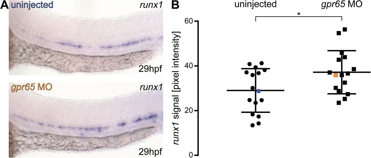
Fig. 3
Gpr65 morphants have significantly increased levels of runx1 mRNA detected by ISH. (A) Representative images of ISH for runx1 in 29 hpf wild-type (blue) and gpr65 MO-injected (orange) embryos, showing the expression in the dorsal aorta. (B) Pixel intensity values of runx1 mRNA in in gpr65 MO-injected embryos (n=16) are significantly higher than in uninjected control siblings (n=16). The coefficients of variation are 34% and 26% for wild-type and morphant groups, respectively. Blue and orange data point correspond to the example images from panel A. The bars represent mean±s.d. *P<0.05 (t-test). The power of the t-test to detect the difference at 0.05 level was 63%.