
Fig. 7
- ID
- ZDB-IMAGE-180705-12
- Genes
- Publication
- Liu et al., 2018 - Lipoprotein lipase regulates hematopoietic stem progenitor cell maintenance through DHA supply
- All Figures
- Figures for Liu et al., 2018
|
Fig. 7
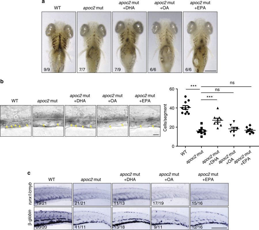
DHA rescues hematopoiesis in apoc2 mutants. a–c apoc2 mutant embryos were injected with free fatty acid docosahexaenoic acid (DHA), oleic acid (OA), or eicosapentaenoic acid (EPA) at 48 hpf. a o-Dianisine staining of 6.3 dpf larvae. b Representative bright field images and quantitative results of blood cell (yellow arrows) count in the caudal vein (outlined with white dashed lines) at 6.3 dpf. Mean ± SEM; n = 10 (WT), n = 8 (apoc2 mut, apoc2 mut + OA, and apoc2 mut + EPA), and n = 9 (apoc2 mut + DHA). ***P < 0.001 (Student’s t test). c In situ hybridization with cmyb/runx1 and β-globin probes. Scale bars, 100 μm in a; 50 μm in b; and 200 μm in c