
|
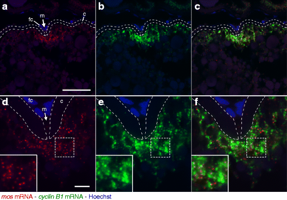
Fig. 3
Double fluorescence in situ hybridization of mos (red) and cyclin B1 (green) mRNAs in zebrafish ovaries. DNA is shown in blue. a-c A zebrafish ovary section showing localization of mos (a) and cyclin B1 (b) mRNAs in a fully grown oocyte. A merged image is shown in (c). The mos and cyclin B1 mRNAs were localized at the animal polar cytoplasm beneath the micropyle (m). fc, follicle cells; c, chorion. d-f High resolution imaging of the oocyte shown in a-c. The insets are enlarged views of the boxed regions. The mos and cyclin B1 mRNAs were distributed in the animal polar cytoplasm of oocyte as different granules. Bars: 50 μm in a-c, 10 μm in d-f