
Fig. 6
- ID
- ZDB-IMAGE-180522-10
- Antibodies
- Publication
- Kimelman et al., 2017 - Regulation of posterior body and epidermal morphogenesis in Zebrafish by localized Yap1 and Wwtr1
- All Figures
- Figures for Kimelman et al., 2017
|
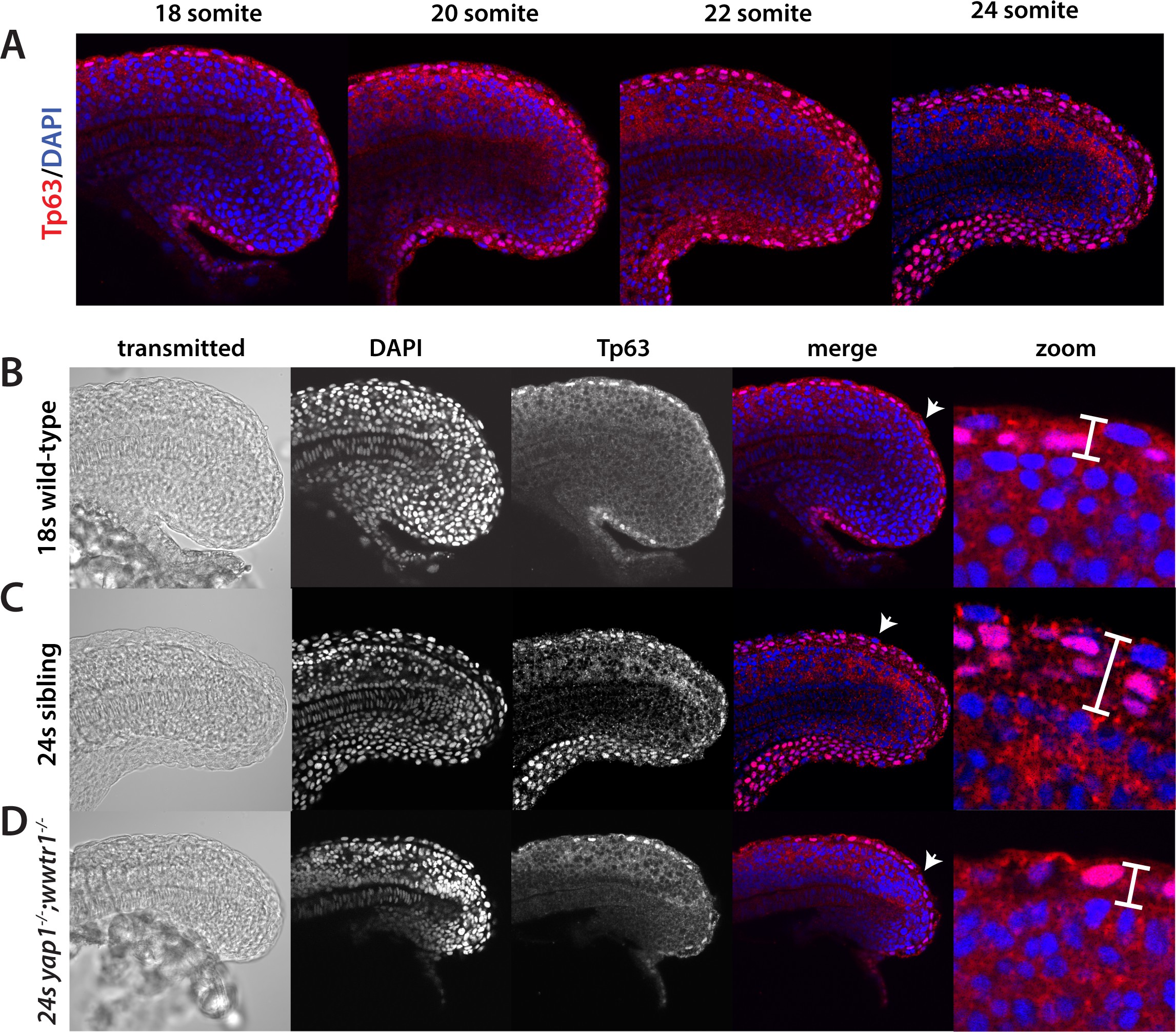
Fig. 6
Tp63 positive cells accumulate at the nascent fin fold in sibling but not mutant embryos.
Embryos were incubated with the anti-Tp63 antibody, and then co-stained with DAPI. (A) Embryos were collected at the indicated stages. Note the increase in Tp63-positive cells on the dorsal and ventral midline as the embryos age. (B–D) The Tp63-positive presumptive epidermis at the 18-somite stage (B) is one layer thick, but increases to multiple layers by the 24-somite stage in sibling (C) but not in yap1;wwtr1 double mutant embryos (D). Confocal images of the midline of the tailbud, with anterior to the left. The zoomed in images in panels B-D are of the dorsal side, and the arrows in the merged images point to the rare presumptive epidermis Tp63-negative cells.