
|
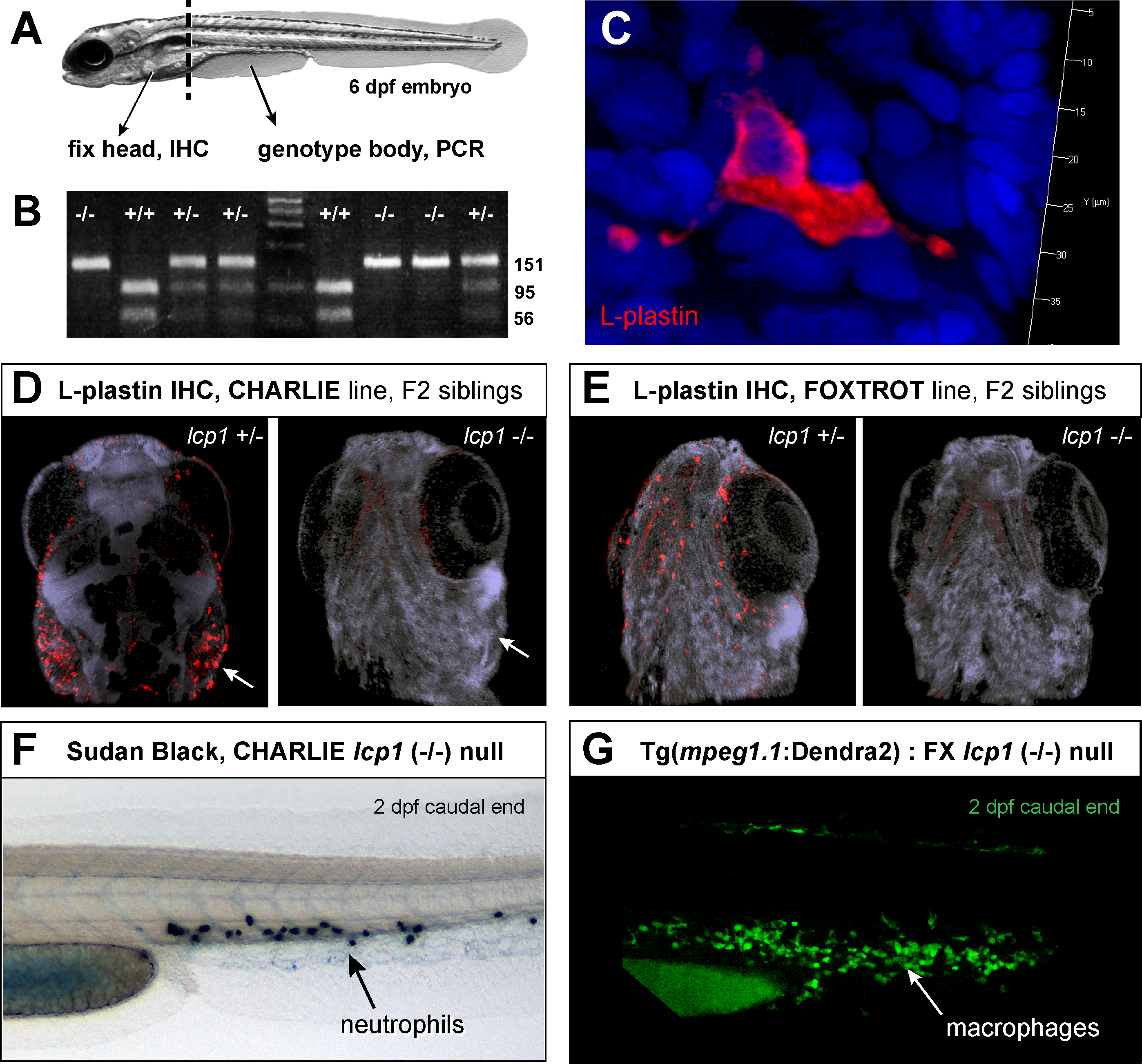
Fig. 5
Leukocyte staining is abolished in lcp1 null animals.
A) Flow chart of the experiment. lcp1 incrosses (+/- x +/-) were raised to 6 dpf and then processed for whole-mount immunostaining (head) and genomic DNA isolation (body). B) Representative genotyping results after genotyping PCR and Bsl1restriction enzyme digest. Homozygous wildtype, homozygous null, and heterozygous siblings are easily distinguished. C) Typical fluorescent immunostaining of a superficial leukocyte in wildtype and heterozygous animals. There is intense signal in the entire cytoplasm, including distant cellular projections; in contrast, the nucleoplasm is dark. D) Heterozygous and null siblings of the Charlie line (CH). In heterozygous fish, leukocytes are stained intensely, particularly in the gill area (arrow). In null fish, no leukocytes are visible. A faint, non-specific staining is present in all embryonic skeletal muscle. E) Heterozygous and null siblings of the Foxtrot line (FX). In the null animal, the LCP1-positive cells are undetectable. F) Null animals have neutrophils as seen in 2 dpf caudal fins stained with Sudan Black. G) Null animals have macrophages, as seen in 2 dpf caudal fins from embryos with green macrophages (Tg(mpeg1.1:Dendra2)). Nuclei are counterstained with DAPI.