Fig. 7
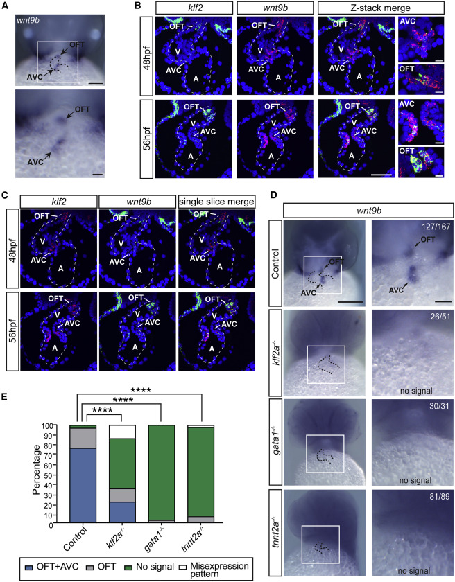
wnt9b Expression in the Developing Zebrafish Heart Is Regulated by Hemodynamic Shear Forces
(A) Whole-mount in situ hybridization for wnt9b in 48 hpf wild-type zebrafish embryos. Dotted lines outline the heart. Scale bars: top, 0.1 mm; bottom, 0.03 mm.
(B and C) In situ hybridization using RNAscope for klf2a and wnt9b in 48 hpf and 56 hpf wild-type zebrafish embryos is shown using merged z stack (B) and single slice (C) confocal analysis. Scale bar represents 100 μm and 10 μm for the magnified images in (B).
(D) In situ hybridization to detect wnt9b expression in the developing AV cushion and outflow tract of 48 hpf wild-type fish (control), silent heart (tnnt2a−/−) mutants that lack blood flow, gata1 mutants (gata1−/−) that experience low shear stress due to low blood viscosity, and klf2a mutants (klf2a−/−) is shown. Dotted lines outline the heart. Scale bars: left, 0.2 mm; right, 0.06 mm.
(E) The percentage of embryos in which wnt9b expression is normal, absent, or mis-expressed in the AV cushion or OFT is shown. The n for each group is indicated in (D). ∗∗∗∗p < 0.0001 using chi-squared analysis.
A, atrium; AV, atrioventricular; OFT, outflow tract; V, ventricle.
Reprinted from Developmental Cell, 43(3), Goddard, L.M., Duchemin, A.L., Ramalingan, H., Wu, B., Chen, M., Bamezai, S., Yang, J., Li, L., Morley, M.P., Wang, T., Scherrer-Crosbie, M., Frank, D.B., Engleka, K.A., Jameson, S.C., Morrisey, E.E., Carroll, T.J., Zhou, B., Vermot, J., Kahn, M.L., Hemodynamic Forces Sculpt Developing Heart Valves through a KLF2-WNT9B Paracrine Signaling Axis, 274-289.e5, Copyright (2017) with permission from Elsevier. Full text @ Dev. Cell

