
|
Fig. 1
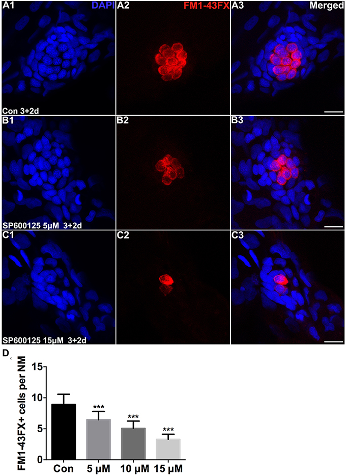
Figure 1. SP600125 reduced the number of FM1−43FX+ cells. (A–C) We treated 3 dpf zebrafish with or without SP600125 for 2 days and subsequently imaged FM1-43FX+ cells (red). Higher magnification of hair cells of the neuromast taken from z-stacks show that (A) hair cells in untreated controls and (B,C) SP600125-treated animals had no observable morphological differences though there were fewer hair cells in the SP600125-treated animals. Nuclei are stained with DAPI and scale bars = 10 μm. (D) The average number of FM1−43FX+ cells per neuromast (NM) in larvae treated with or without SP600125 for 2 days. The first 4 neuromasts along the body, L1–L4, were recorded on one side of each fish. n = 26 neuromasts in control, n = 28 in 5 μM SP600125-treated neuromasts, n = 40 in 10 μM SP600125-treated neuromasts, and n = 28 in 15 μM SP600125-treated neuromasts. One-way ANOVA; FM1−43FX+ cells: F3, 118 = 96.18, p < 0.001. Bars are mean ± SD. ***p < 0.001, highly significant difference when compared to control larvae.