
IMAGE
Fig. 3
- ID
- ZDB-IMAGE-171011-9
- Genes
- Publication
- Morrow et al., 2017 - tbx6l and tbx16 are redundantly required for posterior paraxial mesoderm formation during zebrafish embryogenesis
- All Figures
- Figures for Morrow et al., 2017
Image
|
Figure Caption
Fig. 3
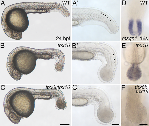
tbx6l; tbx16 double mutants have an enlarged tail bud and lack tail somites. A–C′: Brightfield images of 24-hpf live wild-type (A, A′), tbx16 (spt) mutant (B,B′), and tbx6l; tbx16 double-mutant (C,C′) embryos. tbx6l single-mutant embryos are not shown here as they are indistinguishable from wild-type embryos (see Fig. 1F,G). Black dots in the magnified views (A′–C′) indicate morphologically visible tail somites. D–F: In situ hybridization for msgn1 at the 16-somite stage. Scale bar in A (for A–C) = 250 μm. Scale bar in A′ (for A′–C′) = 100 μm. Scale bar in F (for D–F) = 50 μm.
Figure Data
Acknowledgments
This image is the copyrighted work of the attributed author or publisher, and
ZFIN has permission only to display this image to its users.
Additional permissions should be obtained from the applicable author or publisher of the image.
Full text @ Dev. Dyn.