
Fig. 2
- ID
- ZDB-IMAGE-171004-8
- Genes
- Publication
- Demy et al., 2017 - Trim33 / Tif1-γ is essential for macrophage and neutrophil mobilisation to developmental or inflammatory cues
- All Figures
- Figures for Demy et al., 2017
|
Fig. 2
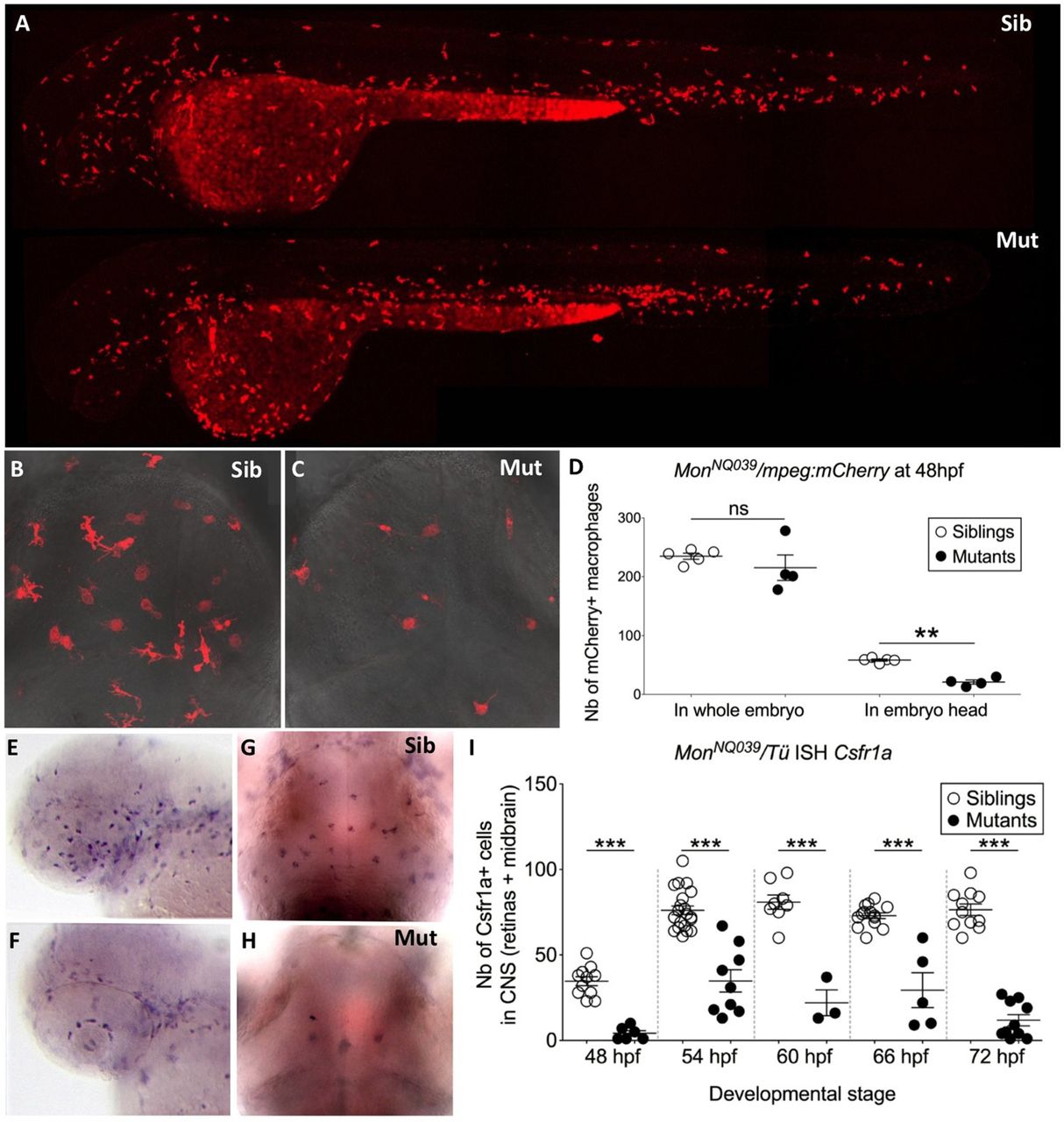
Macrophages are produced normally but do not colonize the CNS in moonshine mutants. (A–C) In vivo images of whole monNQ039Tg(mpeg1:mCherryF) siblings (Sib) (A, upper) and mutants (Mut) (A, lower) at 48 hpf, and dorsal view (rostral up) of the midbrain of siblings (B) and mutants (C) at 72 hpf. (D) Counts (Nb, number) at 48 hpf show that monNQ039Tg(mpeg1:mCherryF) mutant embryos have significantly less mCherry-positive macrophages in the head despite having a similar total number of macrophages to that in the siblings. ISH, whole-mount in situ hybridization. (E–I) Whole mount in situ hybridization for Csfr1a mRNA reveals numerous macrophages in the retina at 48 hpf (E) and midbrain at 72 hpf (G) in the siblings, whereas very few such cells can be detected in these organs in the mutants (F,H). (I) Counting of these cells over time reveals a lower number in the mutant CNS at all stages. Error bars show mean±s.e.m. **P<0.01; ***P<0.001; ns, not significant.