
Fig. 4
- ID
- ZDB-IMAGE-170922-13
- Genes
- Publication
- Sugden et al., 2017 - Genetic dissection of endothelial transcriptional activity of zebrafish aryl hydrocarbon receptors (AHRs)
- All Figures
- Figures for Sugden et al., 2017
|
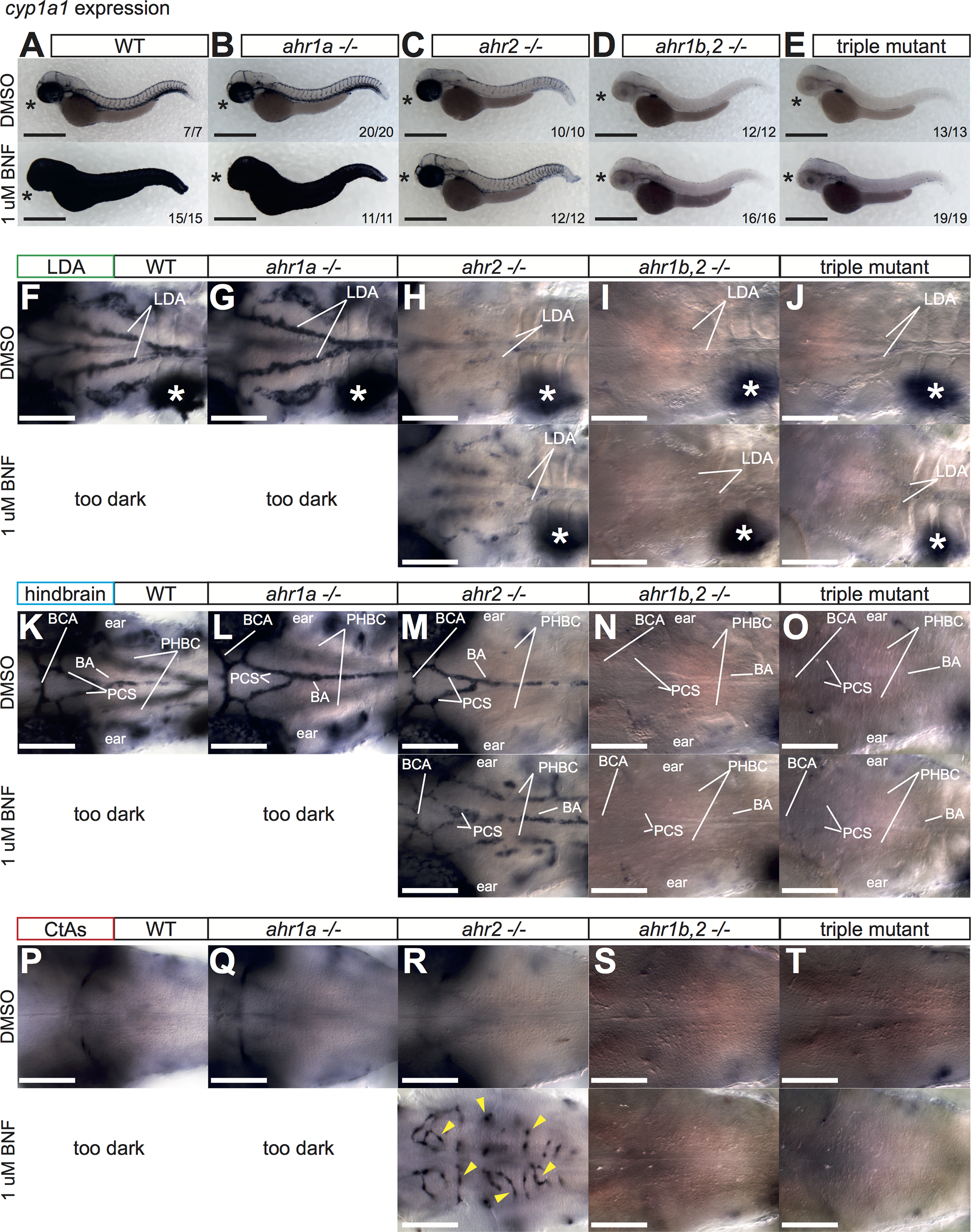
Fig. 4
Regulation of cyp1a1 by AHR.
(A) Overview pictures of cyp1a1 expression at 52 hpf in DMSO or BNF-treated WT (A), ahr1a -/- (B), ahr2 -/- (C), ahr1b,2 -/- (D) and triple AHR mutants (E). ahr1a -/- are indistinguishable from WT. (*) indicates expression in the eye in DMSO and BNF-treated embryos, which is lost only in ahr1b,2 -/- and triple AHR mutants. Note reduction of endogenous vascular cyp1a1 expression in ahr2 mutants (C, upper panels) that is lost in ahr1b,2 -/- and triple AHR mutants (D, E upper panels). ahr2 -/- embryos treated with BNF do not induce cyp1a1 expression in the skin, revealing staining in blood vessels underneath (C, lower panel), which is abolished in ahr1b,2 -/- and triple AHR mutants (D, E, lower panels). Scale bar is 500 um. (F-T) High magnification images of blood vessels of 3 different planes of the head vasculature: the LDA (F-J), the hindbrain blood vessels (K-O) and the hindbrain capillaries (P-T) in all genetic combinations with DMSO and BNF treatment. Note strong dependence of LDA (but not hindbrain arteries, PHBC or CtAs) on ahr2 for full cyp1a1 expression. White asterisk denotes liver. Yellow arrowheads label individual CtAs. Numbers indicate embryos with indicated expression pattern/total embryos of that genotype analyzed. Scale bar is 100 um. Abbreviations–AHR: aryl hydrocarbon receptor, BNF: beta-naphthoflavone, CtA: central artery, CYP: cytochrome p450, DMSO: dimethylsulfoxide, hpf: hours post fertilization, LDA: lateral dorsal aortae, PHBC: primordial hindbrain channel, WT: wildtype.