
IMAGE
Fig. S1
- ID
- ZDB-IMAGE-170706-7
- Genes
- Antibodies
- Publication
- Schall et al., 2017 - Short bowel syndrome results in increased gene expression associated with proliferation, inflammation, bile acid synthesis and immune system activation: RNA sequencing a zebrafish SBS model
- All Figures
- Figures for Schall et al., 2017
Image
|
Figure Caption
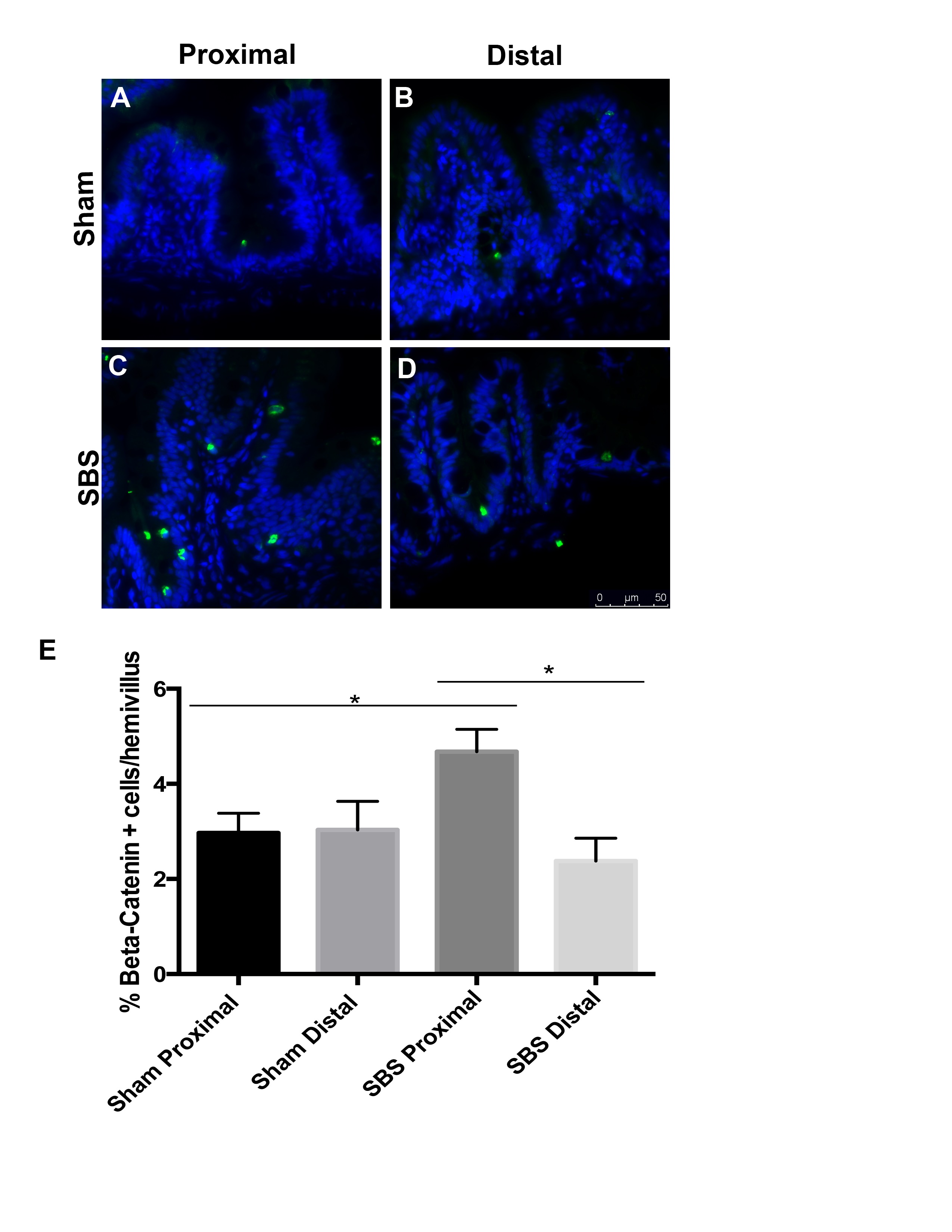
Fig. S1
Increased β –catenin is detected by immunofluorescence in proximal SBS intestine compared to distal SBS and Sham intestine. Immunofluorescent detection of β –catenin identified more positive cells/hemivillus in SBS intestine compared to both the distal limb and sham proximal controls (A–C). Increased β –catenin is noted in the proximal SBS intestine (C; E) compared to distal SBS (D; E p = 0.001) and Sham proximal and SBS proximal intestine (A–C; E p = 0.012). No significant difference in β –catenin is seen between Sham proximal and distal intestine (A–B, E). A–D Scale 50 μm. (TIF 2151 kb)
Figure Data
Acknowledgments
This image is the copyrighted work of the attributed author or publisher, and
ZFIN has permission only to display this image to its users.
Additional permissions should be obtained from the applicable author or publisher of the image.
Full text @ BMC Genomics