
Fig. 5
- ID
- ZDB-IMAGE-170428-30
- Genes
- Publication
- El-Rass et al., 2017 - Disruption of pdgfra alters endocardial and myocardial fusion during zebrafish cardiac assembly.
- All Figures
- Figures for El-Rass et al., 2017
|
Fig. 5
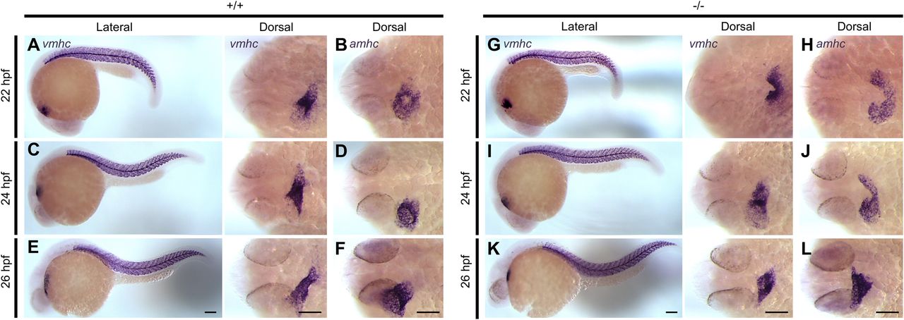
Delay in anterior cardiac fusion disrupts the development of cardiac chambers in pdgfra mutants. Representative images of embryos subjected to whole-mount in situ hybridization with riboprobes against ventricular myosin heavy chain (vmhc) and atrial myosin heavy chain (amhc). In +/+ embryos, the ventricular cells are observed at the apex of the cone (A) and the atrial cells at its base (B) at 22 hpf. At 24 and 26 hpf, the ventricles elongates (C and E), and the atria become cohesive and tubular (D and F). In −/− mutants both the ventricular (G) and atrial (H) regions of the cardiac cone remain incompletely fused at 22 hpf. At 24 to 26 hpf, the anterior portions of the bilateral cardiac fields begin to come into contact; however, the ventricles (I and K) and the atria (J and L) appear shorter and wider. Lateral and dorsal views are shown, with the fish cranium on the left. Scale bar: 100 μm.