
|
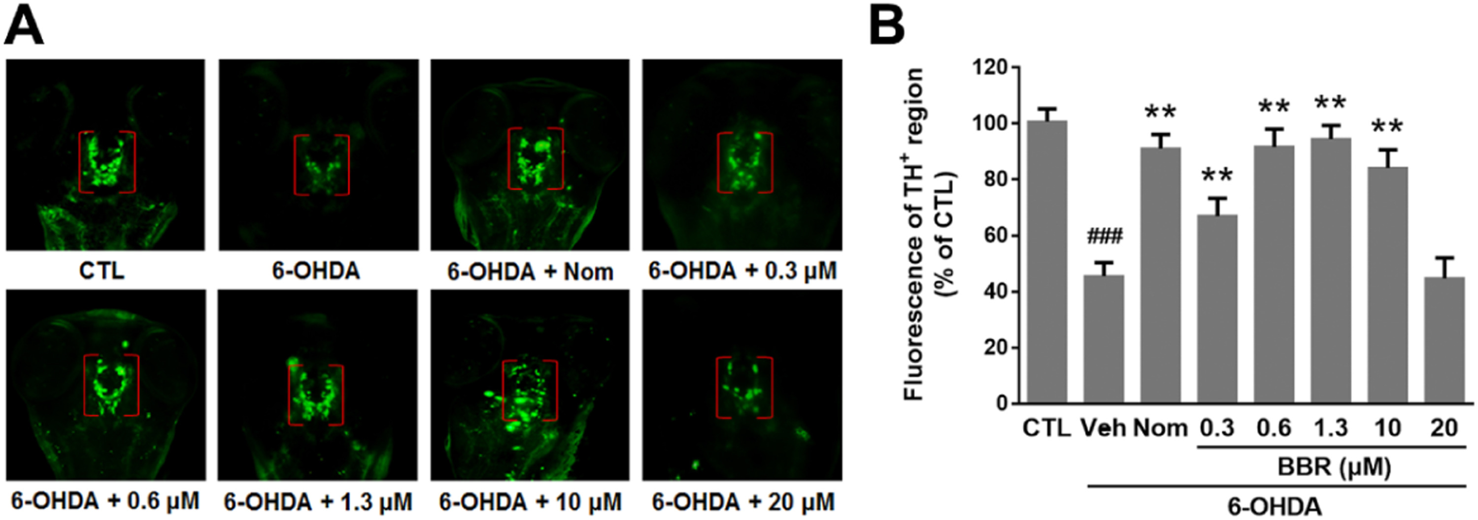
Fig. 5
The effect of BBR on 6-OHDA-induced dopaminergic (DA) neuron loss in zebrafish. Zebrafish embryos at 1 day post fertilization were exposed to different concentrations of BBR or nomifensine (Nom, used as a positive control), and then treated with or without 250 µM 6-OHDA for 48 h. Then zebrafish larvae were fixed for whole-mount immunostaining. (A) Representative morphology of DA neurons in the zebrafish brain indicated by immunostaining with antibody against tyrosine hydroxylase (TH). (B) Statistical analysis of TH+ neuron in each group, 10 fish/group were used. Values represent the mean ± SD (n = 3). Data is expressed as a percentage of the control group. ###P < 0.01 versus control (CTL) group, **P < 0.01 versus vehicle (Veh) + 6-OHDA-treated group.