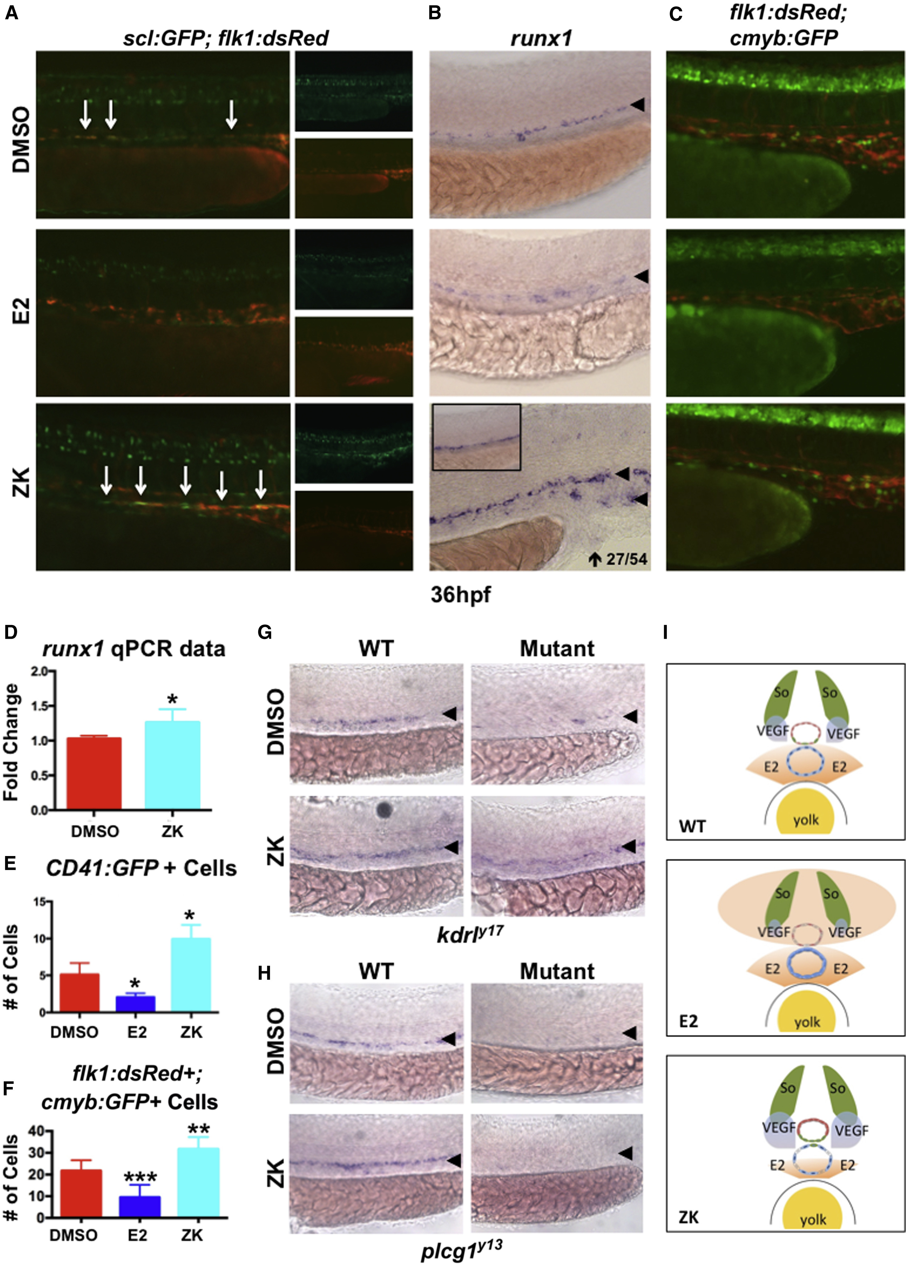
Fig. 7
E2-Mediated VEGF Antagonism Regulates Hemogenic Endothelial Specification
(A) Hemogenic endothelium, as assessed by the dual expression of scl:GFP and flk1:dsRed, was reduced by exogenous E2 treatment but was enhanced following Esr antagonism by ZK (n > 15).
(B) runx1 expression increased (27/54) after estrogen receptor inhibition, with mislocalization to the vein (10/54).
(C) Production of flk1:dsRed+; cmyb:GFP+ HSCs was enhanced by ZK (n ≥ 10).
(D) Quantification of changes in runx1 expression (mean of triplicate experiments ± SEM; one-tailed t test ∗p < 0.05).
(E) Quantification of the number of CD41+ cells after E2 treatment and antagonism (one-tailed t test ∗p < 0.05; error bars indicate SD).
(F) The number of flk1:dsRed+; cmyb:GFP+ HSCs was decreased by E2 and enhanced after ZK treatment (one-tailed t test ∗∗p < 0.01, ∗∗∗p < 0.001; error bars indicate SD).
(G) kdrly17 mutant embryos reveal loss of runx1, which can be partially restored by reduction of E2-mediated VEGF antagonism after ZK treatment (n > 20/condition).
(H) WISH analysis in plcg1y13 mutant embryo shows complete loss of runx1 expression in the presence or absence of ZK (n > 20/condition).
(I) Model: in wild-type embryos (upper panel), opposing gradients of E2 and VEGF specify the arterial/venous boundaries of hemogenic endothelium and subsequent emergence of HSPCs (red circle, artery; blue circle, vein). Excess E2 (middle) disrupts this gradient, further antagonizing VEGF and causing artery, hemogenic endothelium, and HSPCs to fail to form properly. In contrast, inhibition of esr activity (lower panel) allows the range of VEGF regulation to increase, resulting in ectopic HE specification and HSPC production in the vein.

