
Image
|
Figure Caption
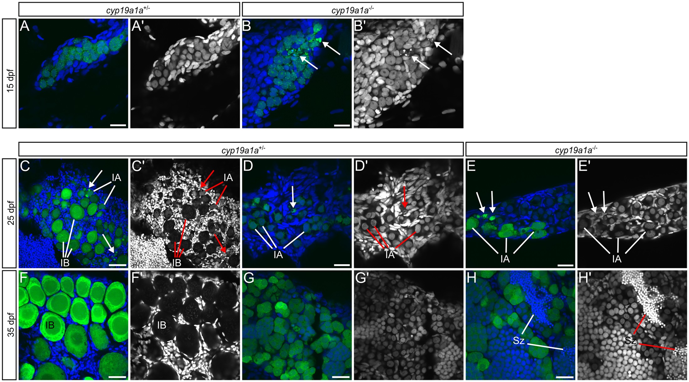
Fig. 4
(A-H′) cyp19a1a heterozygous (A, C, D, F, G) and mutant (B, E, H) gonads at 15 (A-B′), 25 (C-E′), and 35 (F-H′) dpf. Wild-type presumptive ovary (C) and ovary (F). Wild-type presumptive testis (D) and testis (G). Mutant presumptive testis (E) and testis (H). (A-H) Merged images: ziwi:EGFP in green, DNA in blue. (A′-H′) DNA only. Arrows indicate degrading germ cells. IA, stage IA oocyte; IB, stage IB oocyte; Sz, spermatozoa Scale bars: 20 µm (A-E), 50 µm (F -H′).
Figure Data
Acknowledgments
This image is the copyrighted work of the attributed author or publisher, and
ZFIN has permission only to display this image to its users.
Additional permissions should be obtained from the applicable author or publisher of the image.
Full text @ PLoS Genet.