
Fig. 4
- ID
- ZDB-IMAGE-161026-12
- Genes
- Publication
- Deshwar et al., 2016 - Mespaa can potently induce cardiac fates in zebrafish
- All Figures
- Figures for Deshwar et al., 2016
|
Fig. 4
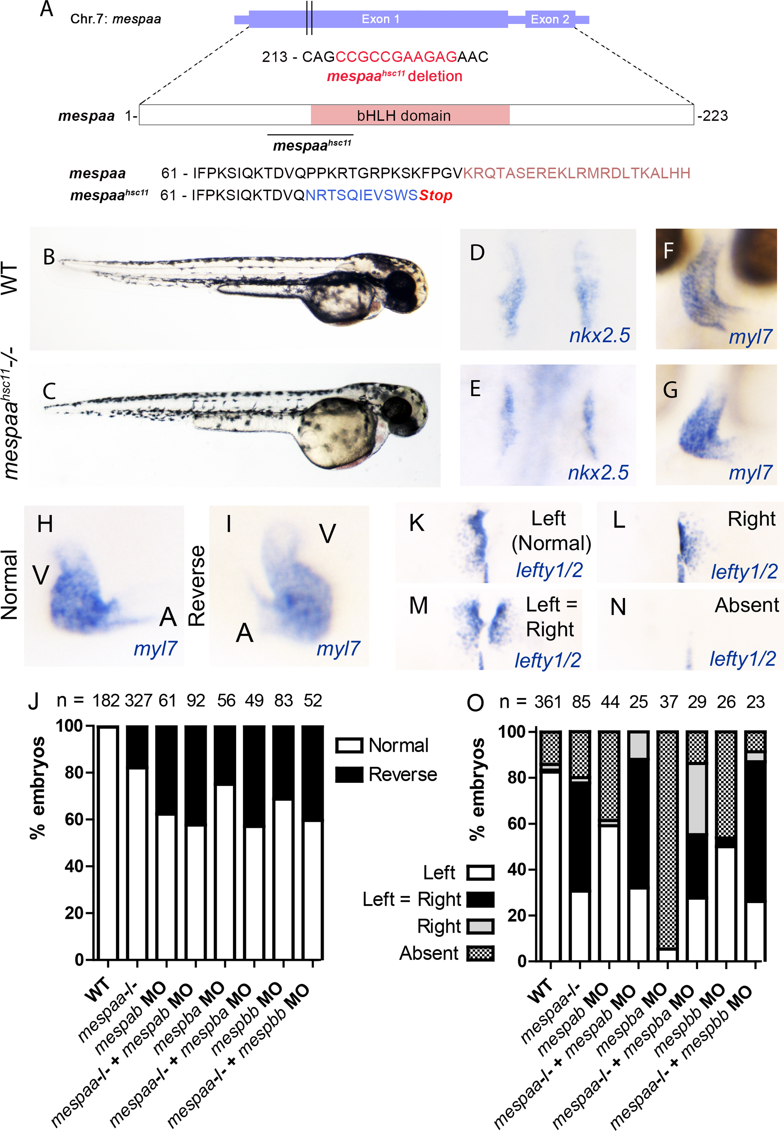
mespaa mutant embryos do not display defects in cardiac specification but exhibit abnormalities in left-right asymmetry. (A) Schematic of the mespaahsc11 null allele that was generated. (B-C) Whole embryo morphology of WT and mespaahsc11 null mutants at 48 hpf. (D-E) nkx2.5 expression in WT and mespaahsc11 null embryos at 16 hpf. (F-G) myl7 expression in WT and mespaahsc11 null embryos at 48hpf. (H-I) Normal and reversed heart looping demonstrated with myl7 expression at 48 hpf. A demarcates the atrium and V indicates the ventricle. (J) Percent of embryos displaying normal or reversed heart looping when deficient for different Mesp family members alone or in combination when compared to WT. (K-N) The four categories of lefty1/2 expression observed in WT and Mesp deficient embryos. (O) Percent of embryos displaying the different lefty1/2 expression patterns in WT and Mesp deficient embryos.
Reprinted from Developmental Biology, 418(1), Deshwar, A.R., Onderisin, J.C., Aleksandrova, A., Yuan, X., Burrows, J.T., Scott, I.C., Mespaa can potently induce cardiac fates in zebrafish, 17-27, Copyright (2016) with permission from Elsevier. Full text @ Dev. Biol.