
Fig. S3
- ID
- ZDB-IMAGE-160927-39
- Genes
- Publication
- Liu et al., 2016 - Fscn1 is required for the trafficking of TGF-β family type I receptors during endoderm formation
- All Figures
- Figures for Liu et al., 2016
|
Fig. S3
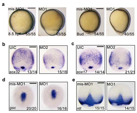
fscn1a morphants exhibit defects in epiboly progression and endoderm formation.
(a) Representative bright-field images of 4 ng fscn1a mis-MO1 and MO1 injected embryos at 8.5 hpf and bud stages. Lateral views with dorsal to the right. (b-c) Knockdown of fscn1a by injection of 25 ng fscn1a MO2 dramatically reduced the expression of the early endodermal markers sox32 (b) and sox17 (c) at the 75% epiboly stage. Panels are shown in dorsal view with anterior to the top. (d-e) The expression of the mesendodermal markers is normal in fscn1a morphants. Embryos injected with mis-MO1 (4 ng) or fscn1a MO1 (4 ng) at the one-cell stage and harvested at the 75%-epiboly stage for in situ hybridization with gsc (d) and ntl (e) probes. Dorsal views with anterior to the top. Scale bar, 200 µm.