
Fig. 3
- ID
- ZDB-IMAGE-160927-31
- Genes
- Antibodies
- Publication
- Liu et al., 2016 - Fscn1 is required for the trafficking of TGF-β family type I receptors during endoderm formation
- All Figures
- Figures for Liu et al., 2016
|
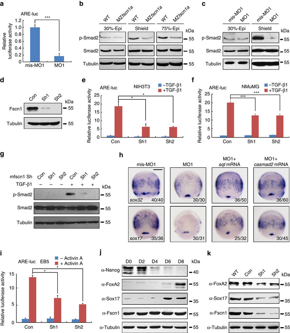
Fig. 3
fscn1a-depletion inhibits Nodal signalling.
(a) Reduction of ARE-luciferase reporter expression in fscn1a morphants. Embryos were co-injected with the reporter plasmids and mis-MO1 or MO1 at the one-cell stage and collected at the 75% epiboly stage for luciferase activity analysis. Data presented as mean with s.d. Student’s t test, n=3, ***P<0.001. (b,c) Wild-type and MZfscn1a mutant embryos (b) and embryos injected with mis-MO1 or MO1 (c) were collected at indicated stages for immunoblotting. (d) The effectiveness of mouse fscn1 shRNAs. NMuMG cells were transfected with 8 µg indicated shRNA plasmids per 100 mm dish and collected 48 h after transfection for western blot analyses. (e-g) NIH3T3 and NMuMG cells transfected with indicated shRNA plasmids (2 µg shRNA plasmids together with or without 0.5 µg reporter plasmids in one well of six-well plate) were treated with TGF-β1 (5 ng mll) for 12 h (e,f) or 3 h (g), and collected for luciferase measurements (e,f) or immunoblotting (g). The relative luciferase activity was the mean with s.d. from three independent biological repeats. Student’s t test, *P<0.05, ***P<0.001. (h) Overexpression of sqt or casmad2 rescues endoderm induction in fscn1a morphants. Embryos were injected with indicated MOs or mRNAs at the one-cell stage and collected at the 75% epiboly stage for in situ hybridization with sox32 and sox17 probes. Injection doses: mis-MO1, 4 ng; MO1, 4 ng; sqt mRNA, 1 pg; casmad2 mRNA, 100 pg. Scale bar, 200 µm. (i) EB5 cells transfected with ARE-luciferase reporter together shRNA plasmids and treated with or without 25 ng ml-1 Activin A for 12 h before collected for luciferase assays. Data presented as mean with s.d. Student’s t test, n=3, *P<0.05. (j) Western blots for the expression of Nanog, FoxA2, Sox17 and Fscn1 in samples taken at specified time points during embryoid body formation. The expression of Tubulin was examined as loading control. (k) Fscn1 knockdown disturbs the formation of endodermal cell lineage during embryoid body differentiation. Embryoid bodies on day 8, derived from EB5 cells infected with the indicated lentiviral shRNAs, were collected for western blots with indicated antibodies.