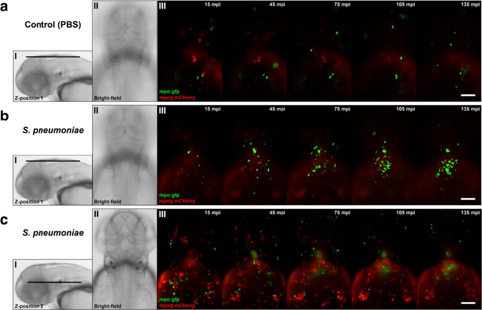
Fig. 8
Time-lapse fluorescence imaging of Streptococcus pneumoniae-infected zebrafish embryos via the hindbrain ventricle at 2 days post fertilization. a-c Live fluorescence images of double-labelled Tg(mpx:GFP) i114 /Tg (mpeg1:mCherry) gl23 zebrafish embryos (green fluorescent neutrophils, red fluorescent macrophages). Dorsal view of the head region after injection with a PBS or b, c S. pneumoniae D39 with the corresponding Z-position (I) and bright-field image (II). Images b and c were acquired from the same zebrafish embryo at different positions. b (III) After injection of pneumococci in the hindbrain ventricle, green fluorescent neutrophils migrate in increasing numbers to the site of infection compared to a (III) PBS-injected zebrafish embryos. Macrophages were not observed in the subarachnoid space during b early pneumococcal infection of the hindbrain ventricle but c (III) remain localized in regions below the subarachnoid space. Scale bars, 100 µm

