
Image
|
Figure Caption
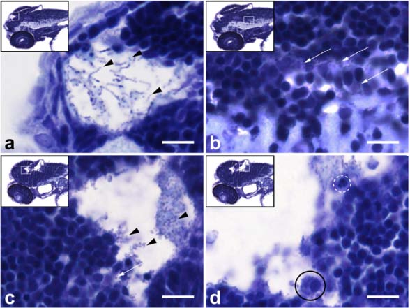
Fig. 4
Histopathological analysis of Streptococcus pneumoniae-infected zebrafish embryos via the hindbrain ventricle at 2 days post fertilization. a, b Sagittal section of the head region showing bacteria (arrow heads) in a the subarachnoid space and b brain parenchyma at 12 h post injection (hpi). c, d Sagittal section at 24 hpi showing increased amount of bacteria in the subarachnoid space (arrow heads) and disruption of the ventricular lining with bacterial infiltration (arrow) in c and a neutrophil (dotted circle) and a phagocytosing macrophage (circle) in d. Scale bars, 10 µm
Acknowledgments
This image is the copyrighted work of the attributed author or publisher, and
ZFIN has permission only to display this image to its users.
Additional permissions should be obtained from the applicable author or publisher of the image.
Full text @ J Neuroinflammation