
Image
|
Figure Caption
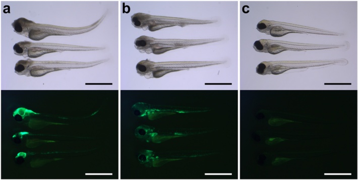
Fig. 2
Bright-field images with corresponding fluorescent images of 2 day-post-fertilization zebrafish embryos infected by different routes. a, b Lateral view of zebrafish embryos injected in the a hindbrain ventricle or b in the caudal vein. c Non-injected control embryos. Please note that there is usually some background fluorescence observed in the yolk. All embryos were infected with 400 CFU of Streptococcus pneumoniae D39 (HlpA-GFP) and imaged at 48 h post injection. Scale bars, 500 µm
Acknowledgments
This image is the copyrighted work of the attributed author or publisher, and
ZFIN has permission only to display this image to its users.
Additional permissions should be obtained from the applicable author or publisher of the image.
Full text @ J Neuroinflammation