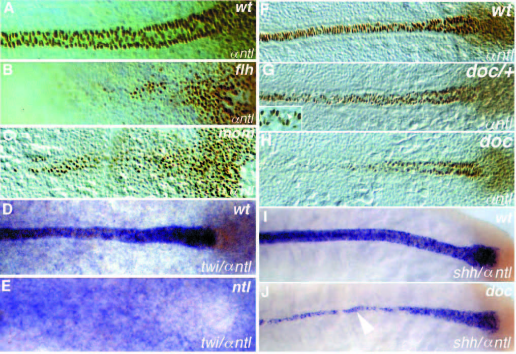
Fig. 3
Dorsal views of (A-E) tailbud stage and (F-J) 10-somite stage embryos. (A-C, F-H) Whole-mount Ntl antibody staining; (D,E) twi in situ hybridization (blue) and Ntl antibody staining (brown); (I, J) shh in situ hybridization (blue) and Ntl antibody staining (brown). Anterior is to the left. (A,D,F,I) Wild type, (B) flhtk241, (C) momth211, (E) ntltc41, (G) heterozygous and (H, J) homozygous doctt258 embryos. Ntl expression in the axial mesoderm is reduced in (B) flh and (C) mom embryos at the tailbud stage. Expression of twi in (D) wild-type and (E) ntltc41 embryos, note that expression in the notochord is not detectable. (G) In heterozygous doctt258/+ embryos, Ntl is not detectable in a few nuclei of the notochord (arrowhead in the enlargement). In (J) doctt258 embryos notochord expression of shh (blue) is only detectable in notochord cells which also express Ntl (brown, compare to Ntl expression in H). In regions to the left of the arrowhead (anterior), shh expression is restricted to the floor plate.

