Fig. 5
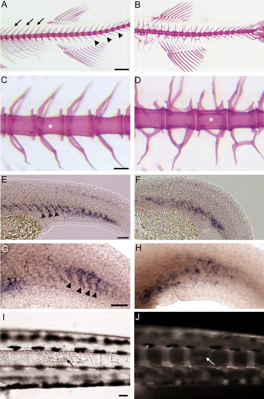
Vertebral segmentation in fused somite (fss) mutant fish. (A-D) Alizarin red staining of 30 dpf larvae. (A) In wild-type larvae, neural (arrows) and haemal (arrowheads) arches extend from each centrum in a regular manner. (B) As reported previously (van Eeden et al., 1996), defects in the position and projection of the neural and haemal arches are conspicuous in fss embryos. (C,D) Higher magnification of A and B, respectively. In wild-type (C) and fss (D) embryos, centra (asterisk) are of uniform size and no fusions or abnormalities are seen in fss centra. (E,F) Pax9 expression in sclerotome of wild-type and mutant embryos at 24 hours post-fertilization (hpf). (E) Wild-type expression in discrete cell clusters, one cluster per somite (arrowheads). (F) In fss mutants, expression is not segmented. (G,H) Smad1 expression in sclerotome of wild-type and fss embryos at 36 hpf. (G) Similar to Pax9, Smad1 expression is seen in one cell cluster per somite (arrowheads). (H) In fss mutants the segmented pattern of Smad1 expression is disrupted. (I,J) Ablation of notochord cells (arrow) in 4-dpf fss embryos prevents development of centra (8 embryos). (I) DIC image of 12-dpf embryo indicating wound site and local cell reorganisation resulting from ablation (arrow). (J) Bone staining of embryo in I showing absence of centrum (arrow indicates the probable anterior centrum boundary) after ablation. Scale bars: A,B, ~500 µm; C,D, ~100µ m; E,F, ~20 µm; G,H, ~20 µm; I,J, ~100 µm.

