
Fig. 3
- ID
- ZDB-IMAGE-160708-39
- Genes
- Antibodies
- Publication
- Wu et al., 2014 - Mediator subunit 12 coordinates intrinsic and extrinsic control of epithalamic development
- All Figures
- Figures for Wu et al., 2014
|
Fig. 3
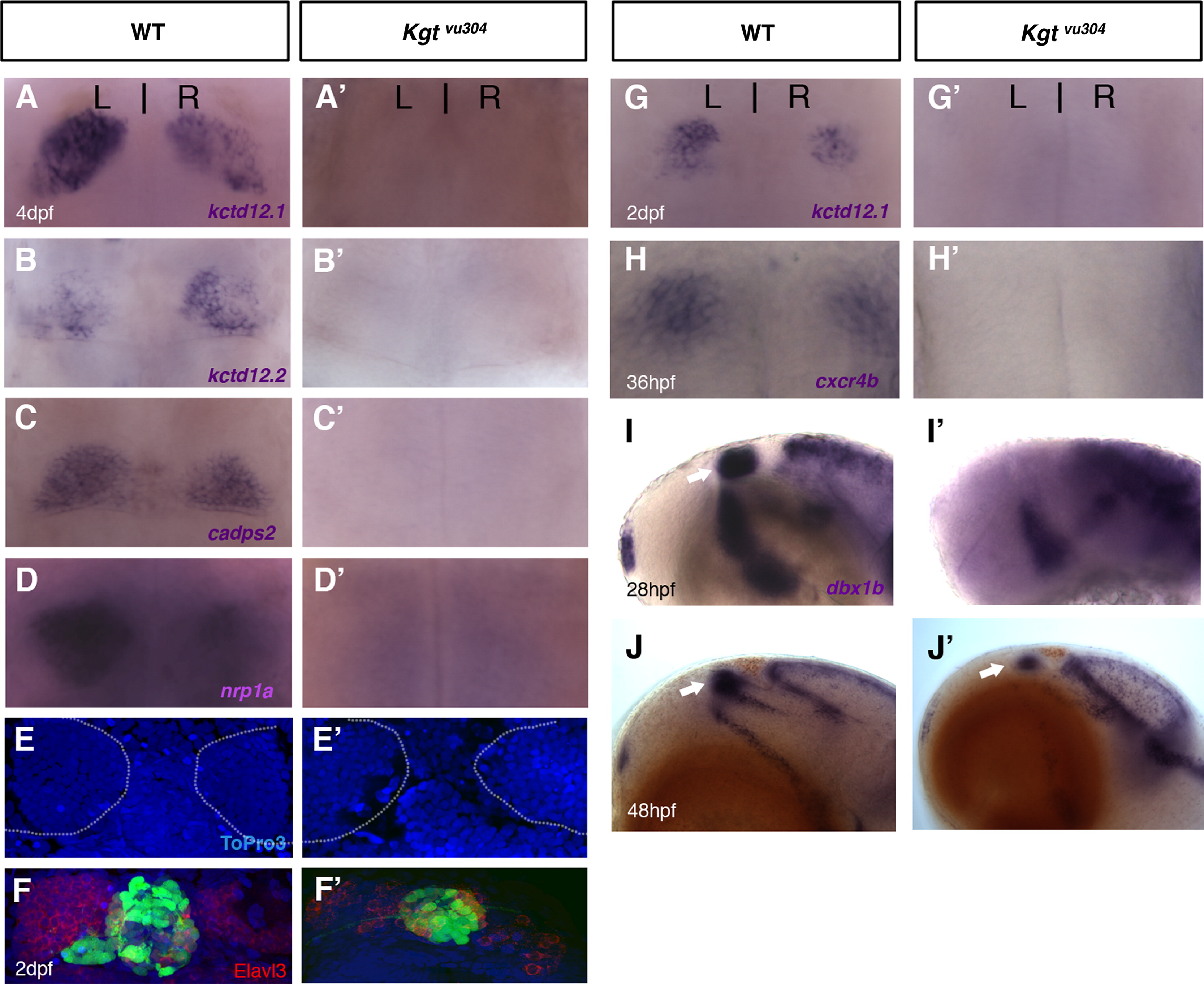
The med12/kgt mutant shows defects in habenular development. The habenular differentiation markers, kctd12.1, kctd12.2, nrp1a, and cadps2 were all absent in med12 mutants at 4 dpf (A-D′) and 2 dpf (G, G′), while the habenular cells were still present in the dorsal diencephalon in med12 mutants as shown by ToPro3 nuclear staining at 4 dpf (E, E′; habenular nuclei were outlined in white; the dark shadow on the left habenula in E′ is due to an overlying pigment cell). Fewer Elavl3-expressing neurons were observed (F, F′; at 2 dpf) and the expression of cxcr4b was significantly reduced in med12 mutants when compared to WT (H, H′; at 36 hpf). In med12 mutants, the presumptive habenular progenitor marker, dbx1b, was absent early (lateral views, at 28 hpf; I, I′) but reappeared in later stages (at 48 hpf, J, J′).
Reprinted from Developmental Biology, 385(1), Wu, S.Y., de Borsetti, N.H., Bain, E.J., Bulow, C.R., and Gamse, J.T., Mediator subunit 12 coordinates intrinsic and extrinsic control of epithalamic development, 13-22, Copyright (2014) with permission from Elsevier. Full text @ Dev. Biol.