
Fig. 7
- ID
- ZDB-IMAGE-160701-35
- Genes
- Publication
- Huang et al., 2013 - Sequential effects of spadetail, one-eyed pinhead and no tail on midline convergence of nephric primordia during zebrafish embryogenesis
- All Figures
- Figures for Huang et al., 2013
|
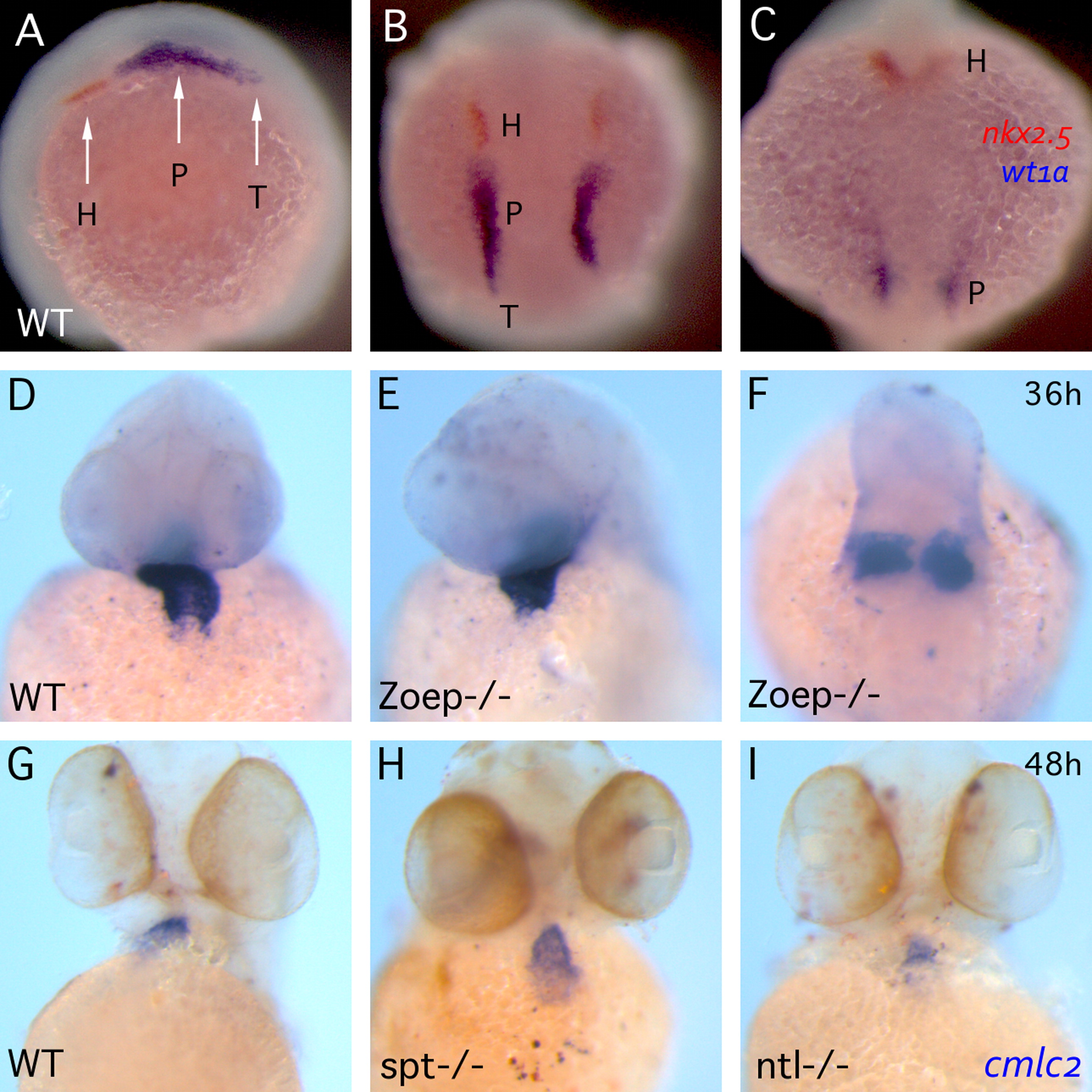
Fig. 7
PGP and cardiac midline convergence. ((A)-(C)) Transiency of PGP neighboring tissue. Double in situ of nkx2.5 (orange) and wt1a (blue) at 12 ((A)-(B)) and 18 hpf (C). (A) The image is lateral view anterior to the right. ((B)-(C)) are dorsal view anterior to the top and same magnification. (H) heart primordium; (P) pronephric glomerular primordium; (T) tubule primordium. ((D)-(I)) Whole-mount in situ hybridization of cmlc2 demonstrating cardiac morphogenetic phenotypes at 36 ((D)-(F)) and 48 hpf ((G)-(I)) in WT ((D) and (G)), Zoep-/- ((E) and (F)), spt-/- (H) and ntl-/- (I) embryos. ((D)-(I)) The images are same magnification and anterior to the top; ((A) and (B), (D)-(F)) are ventral view and (C) is dorsal view.
Reprinted from Developmental Biology, 384(2), Huang, C.J., Wilson, V., Pennings, S., MacRae, C.A., and Mullins, J., Sequential effects of spadetail, one-eyed pinhead and no tail on midline convergence of nephric primordia during zebrafish embryogenesis, 290-300, Copyright (2013) with permission from Elsevier. Full text @ Dev. Biol.