
Fig. 6
- ID
- ZDB-IMAGE-160701-34
- Genes
- Publication
- Huang et al., 2013 - Sequential effects of spadetail, one-eyed pinhead and no tail on midline convergence of nephric primordia during zebrafish embryogenesis
- All Figures
- Figures for Huang et al., 2013
|
Fig. 6
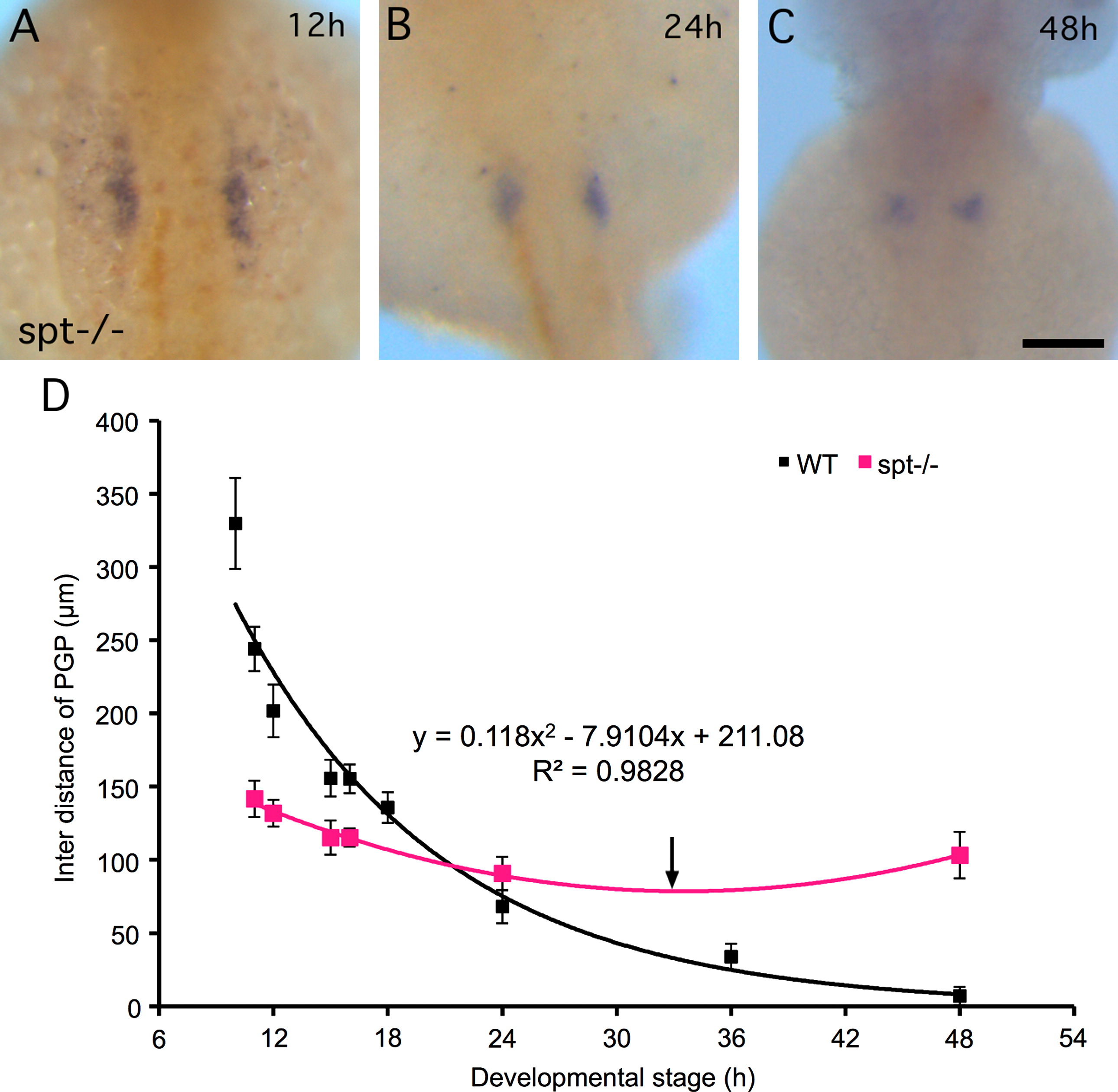
PGP midline convergence phenotypes in spt-/-embryos. ((A)-(C)) Whole-mount in situ hybrydization of wt1a and myoD was stained blue and orange, respectively in 12 (A), 24 (B) and 48 hpf (C) spt-/- embryos. The scale bar indicates 100 µm; ((A)-(C)) are at the same magnification. All images are dorsal view anterior to the top. (D) Quantification of convergence morphogenetic movements in spt-/- embryos and comparison with WT. The inter distance of PGP was measured at 11 (n=28), 12 (n=32), 15 (n=31), 24 (n=9), 48 (n=15) hpf. The P values of t-test for all 5 stages are smaller than 0.005. The curve fitting (pink) and R-squared value are displayed on the right-hand corner. A turning point (arrow) to divergence movement at 33.5 hpf is generated from an approximation of the stage that has the minimum inter distance of PGP. Error bars represent SD.
Reprinted from Developmental Biology, 384(2), Huang, C.J., Wilson, V., Pennings, S., MacRae, C.A., and Mullins, J., Sequential effects of spadetail, one-eyed pinhead and no tail on midline convergence of nephric primordia during zebrafish embryogenesis, 290-300, Copyright (2013) with permission from Elsevier. Full text @ Dev. Biol.