
Fig. 3
- ID
- ZDB-IMAGE-160513-11
- Genes
- Publication
- Zhou et al., 2015 - Gmnc Is a Master Regulator of the Multiciliated Cell Differentiation Program
- All Figures
- Figures for Zhou et al., 2015
|
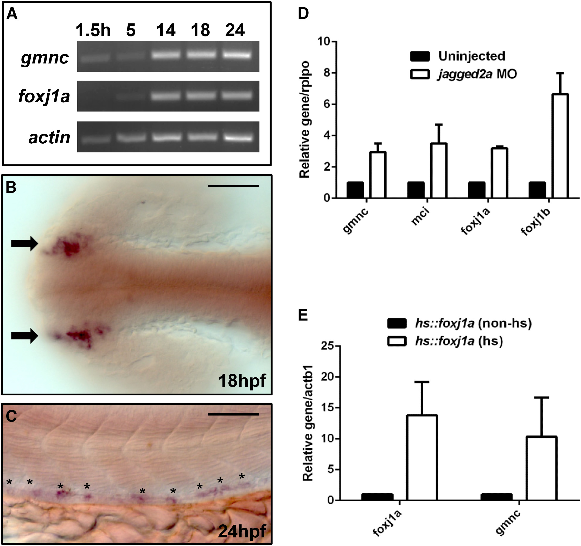
Fig. 3
gmnc Is Expressed in the Kidney Tubules and Is Modulated by Notch Signaling and Foxj1a Activation
(A) End-point PCRs on cDNA extracted from 1.5, 5, 14, 18, and 24 hpf embryos, respectively, revealed that whereas the maternal contribution of the foxj1a transcript is minimal (1.5 hr), the gmnc transcript is maternally deposited. actin-b1 (actb1) was used as the loading control.
(B and C) Representative staining from in situ hybridization using a probe against gmnc showed that the gene is expressed in the developing nasal placodes (arrows) at 18 hpf (B) and is also expressed in a spotted pattern in the mid-section of the kidney ducts (asterisks) at 24 hpf (C). Scale bars, 50 µm.
(D) qPCRs were performed on uninjected or jagged2a MO-injected embryos at 24 hpf. Similar to the ciliogenic genes like foxj1a, foxj1b, and mcidas (mci), whose levels increased in the N-deficient background, gmnc transcript levels also increased moderately (by ~3-fold). Expression levels in the uninjected condition were arbitrarily assigned a value of 1. rplpo was used as an internal (loading) control. Error bars represent the SEM from two independent experiments.
(E) qPCRs were performed on non-heat-shocked or heat-shocked Tg(hsp70::foxj1a) embryos. The heat-shock treatment induced the foxj1a transgene level by ~15-fold, and the foxj1a overexpression induced the gmnc level by ~10-fold. Expression levels in the non-heat-shocked condition were arbitrarily assigned a value of 1. actin-b1 (actb1) was used as an internal (loading) control. Error bars represent the SEM from three independent experiments.