
|
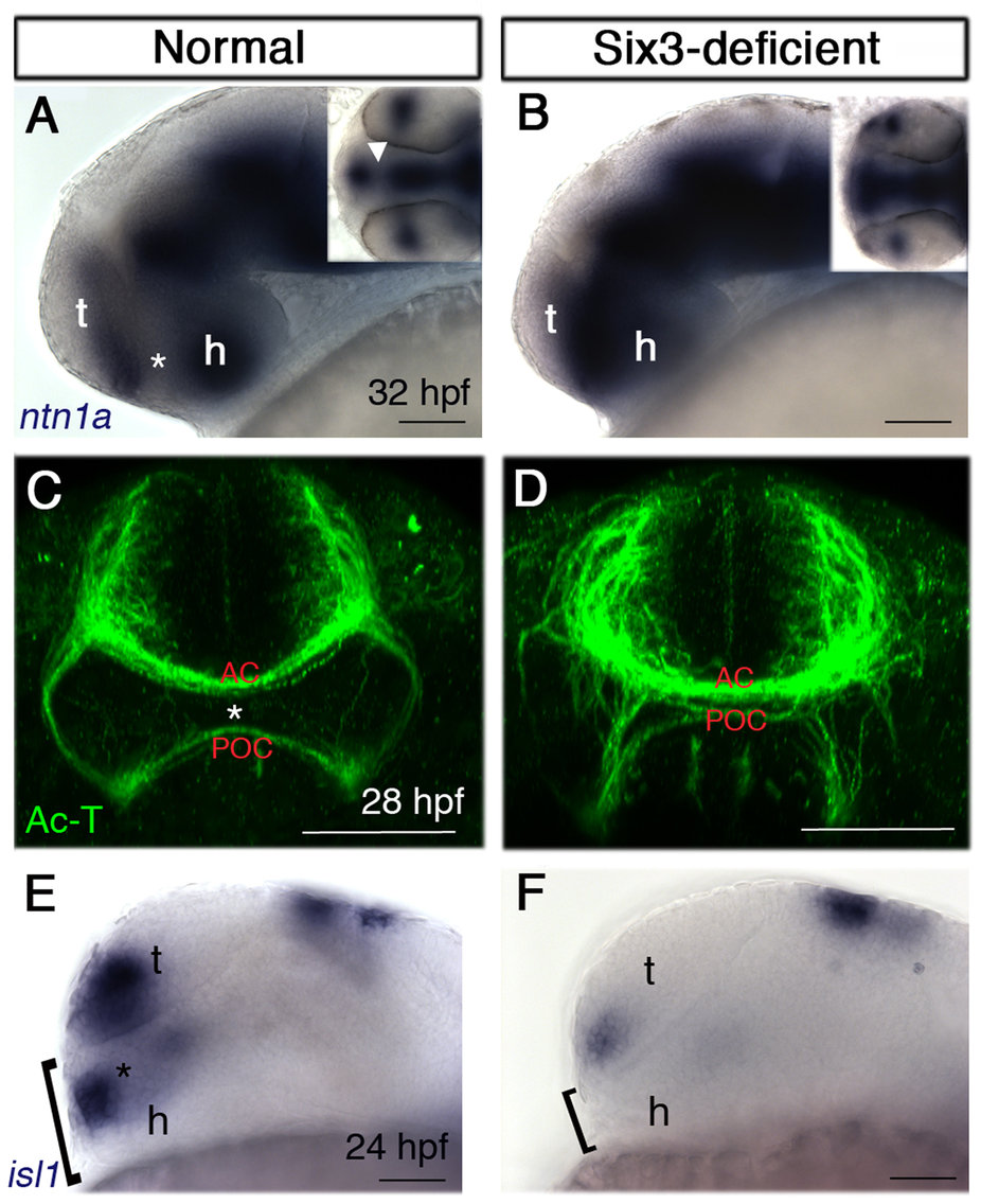
Fig. 6
The POA is severely reduced in Six3-deficient embryos.
(A,B) ntn1a expression at 32 hpf in normal (A) and Six3-deficient (B) embryos. Insets are dorsal views of the same embryos. Asterisk and arrowhead (inset) in A mark the ntn1a-negative POA. (C,D) Ac-T immunohistochemistry at 28 hpf in normal (C) and Six3-deficient (D) embryos. Asterisk in (C) marks the POA. (E,F) isl1 expression at 24 hpf in normal (E) and Six3-deficient (F) embryos. Brackets mark the dorsoventral dimension ventral to the anteriormost telencephalon. AC, anterior commissure; POA, preoptic area; POC, post-optic commissure; t, telencephalon; h, hypothalamus. (A,B,E,F) are lateral views, anterior to the left; (C,D) are frontal views. Scale bars are 50 µm.