
Fig. 6
- ID
- ZDB-IMAGE-160315-17
- Genes
- Antibodies
- Publication
- Govindan et al., 2016 - Cx43-Dependent Skeletal Phenotypes Are Mediated by Interactions between the Hapln1a-ECM and Sema3d during Fin Regeneration
- All Figures
- Figures for Govindan et al., 2016
|
Fig. 6
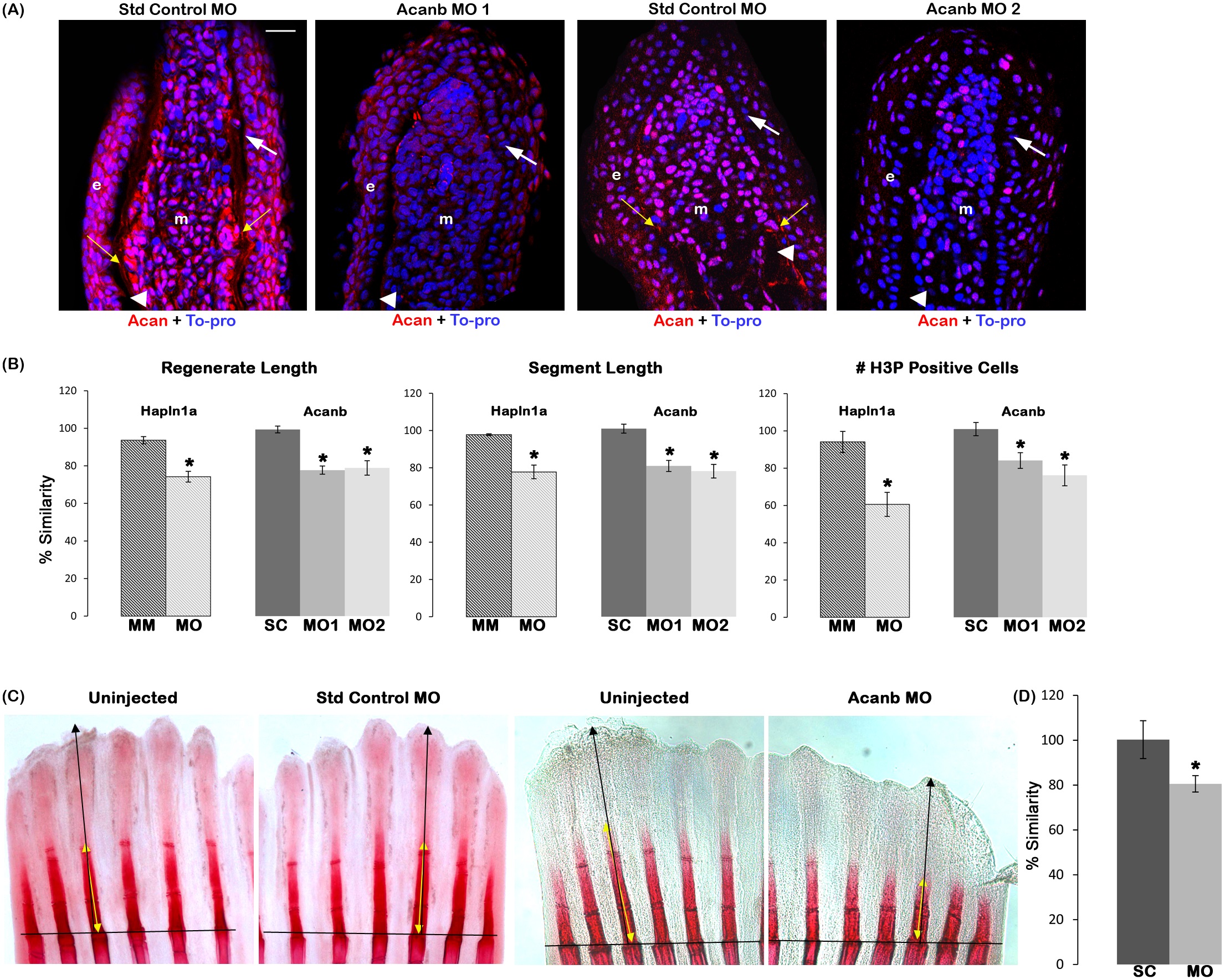
Reduced Acan contributes to Hapln1a knockdown phenotypes.
(A) Longitudinal section of fin rays treated with standard control morpholino (Standard control MO) and acanb ATG-blocking morpholino (Acanb MO1) or splice blocking morpholino (Acanb MO2). Immuno-staining for Acan (red) and counterstained for nuclei with To-pro (blue). Compared to the standard MO treated fins, Acanb MO treated fins show reduced staining for Acan. White arrow identifies the basal layer of epithelium; yellow arrow identifies Acan staining in the lepidotrichia, arrow head identifies the bone; m, mesenchyme; e, epithelium. Scale bar represents 20 µm. (B) Bar graph shows that regenerate length, segment length and cell proliferation are significantly reduced upon acanb knockdown using both MO1 and MO2. The mean of percent similarity for the MO treated experimental group and the corresponding control group were estimated and compared. Statistical significance was determined using the student′s t-test (P<0.05) and the error bars represent standard error of mean. The hashed bars indicate the extent of hapln1a knockdown effect compared to acanb knockdown. (C) Representative alizarin red stained fins showing extent of bone calcification. The black line indicates the amputation plane. (D) The extent of mineralization was calculated as the ratio of the zone of mineralization (extent of detectable alizarin red staining length) to the total regenerate length. The mean of percent similarity for the MO treated experimental group and the corresponding control group were estimated and compared, and the statistical significance between the groups was determined using two tailed unpaired student′s t-test (P<0.05) and the error bars indicate the standard error of mean.