
Fig. 3
- ID
- ZDB-IMAGE-150629-8
- Genes
- Publication
- Rossi et al., 2015 - The SLC7A7 Transporter Identifies Microglial Precursors prior to Entry into the Brain
- All Figures
- Figures for Rossi et al., 2015
|
Fig. 3
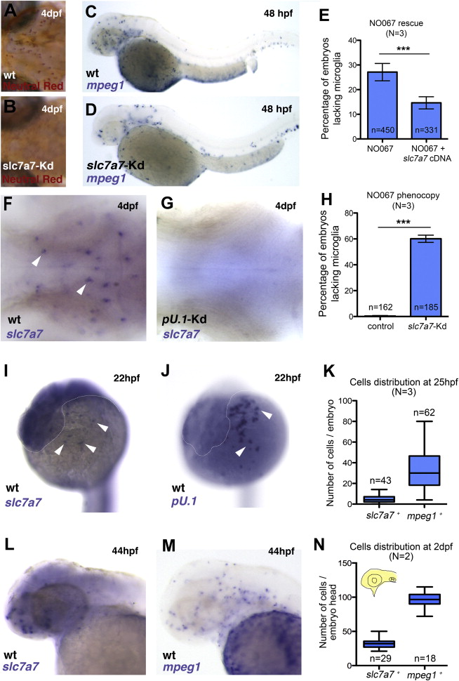
Slc7a7 Is Expressed in Microglia and a Monocyte Sub-lineage at Early Developmental Stages
(A and B) Neutral red staining shows a representative wild-type (A) and slc7a7 morphant (B) embryo at 4 days post fertilization, with a lack of microglia in the slc7a7 morphant brain.
(C and D) WISH for mpeg1 in a wild-type (C) and a slc7a7 morphant (D) embryo at 2 days post fertilization. Slc7a7 morphants lack macrophages in the brain and in the eyes.
(E) Rescue of the NO067 phenotype. Percentages of homozygous NO067 embryos lacking microglia at 4 days post fertilization, with or without expression of the UAS::slc7a7 cDNA, are given. Error bars show SEM. p < 0.001, Fisher’s exact test.
(F and G) WISH for slc7a7 in the brain of a wild-type (F) and a pU.1 morphant (G) embryo at 4 days post fertilization (dorsal views). Slc7a7+ cells are absent in pU.1 morphants, indicating that they have a myeloid origin. White arrowheads indicate slc7a7+ cells.
(H) Phenocopy of the NO067 phenotype. Percentages of 4-day-post-fertilization embryos lacking microglia in control and slc7a7 morphant embryos are given. Error bars show SEM. p < 0.001, Fisher’s exact test.
(I and J) WISH for slc7a7 (I) and pU.1 (J) in a wild-type embryo at 22 hpf. White arrowheads indicate slc7a7+ and pU.1+ cells, respectively. Dotted line indicates the shape of the head.
(K) Numbers of slc7a7+ cells and mpeg1+ cells at 25 hpf are given.
(L and M) WISH shows 2-day-post-fertilization wild-type embryos stained with slc7a7 (L) and mpeg1 (M).
(N) Numbers of slc7a7+ cells and mpeg1+ cells at 2 days post fertilization are given.
N, number of experiments; n, number of embryos. See also Figure S4.