Fig. 1
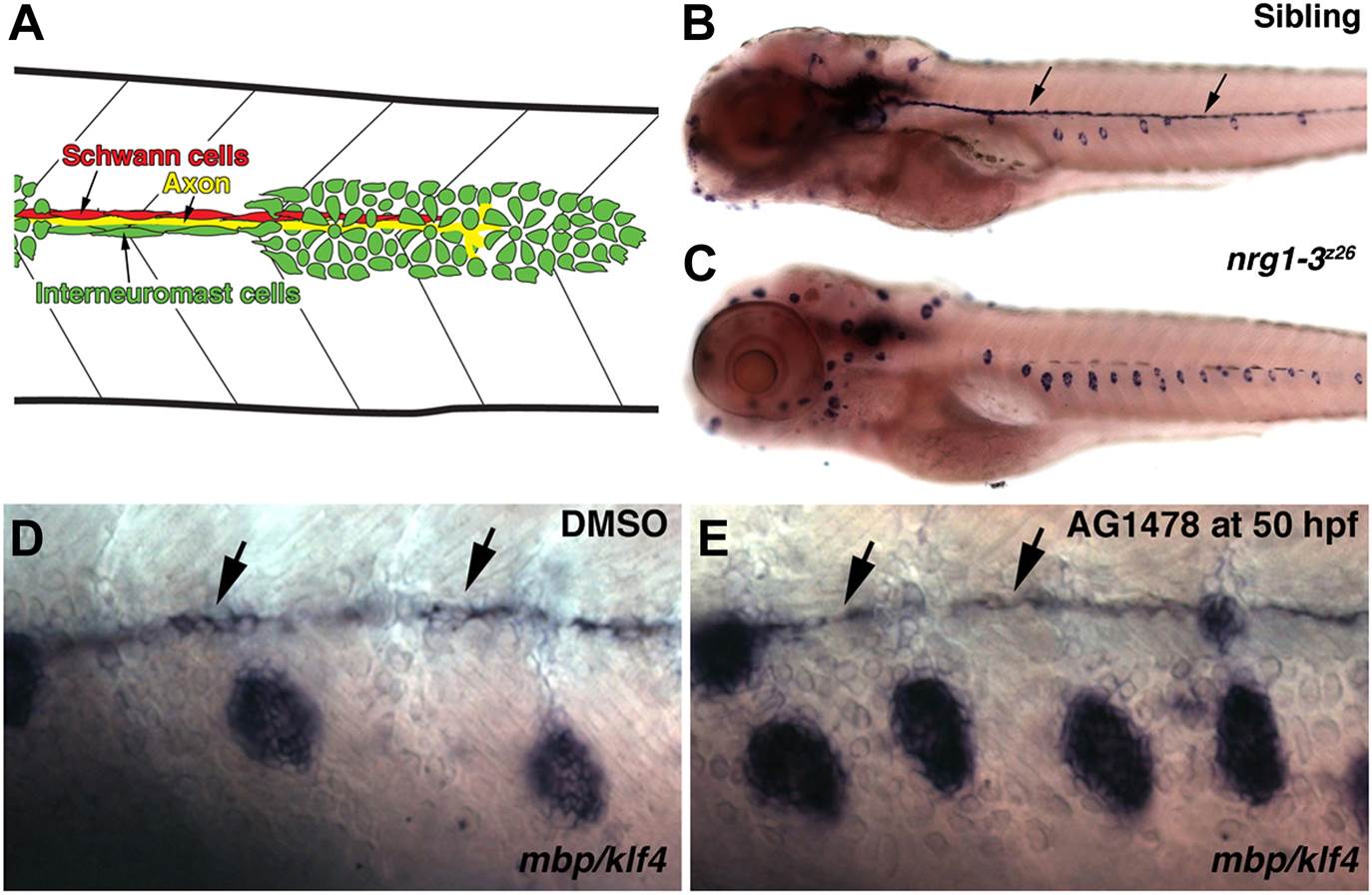
Illustration of cell types in the migrating lateral line.
(A) As the primordium migrates it deposits neuromasts and a chain of interneuromast cells (green cells). Pioneer axons (yellow line) of the posterior lateral line ganglion grow out with the primordium. Schwann cells (red cells) migrate and proliferate along axons. nrg1-3z26 mutants and pharmacological inhibition of ErbB signaling mimics the erbb phenotype. (B–E) Double in situ hybridization was performed to label Schwann cells with myelin basic protein (mbp) and neuromasts with klf4 at 5 dpf. (B) Control siblings with Schwann cells (arrows) along the lateral line nerve and normal neuromast number. nrg1-3z26 mutants mimic erbb2 and erbb3b mutants in that they lack Schwann cells along the lateral line and have increased neuromast number (C). The brown cells along the midline in both sibling and nrg1-3z26 are pigment cells. (D and E) Double in situ hybridization for mbp and klf4 in DMSO or AG1478 treated larvae from 50 hpf. Compared to DMSO treatment (D), increased neuromasts are seen in AG1478 treated larvae (E). mbp expression along the midline shows that Schwann cells (arrows) are still present at 5 dpf when AG1478 was given at 50 hpf (E), compare to DMSO treated (D).

