
Fig. 3
- ID
- ZDB-IMAGE-150427-67
- Genes
- Publication
- Yuan et al., 2015 - Sumoylation of CCAAT/enhancer-binding protein α is implicated in hematopoietic stem/progenitor cell development through regulating runx1 in zebrafish
- All Figures
- Figures for Yuan et al., 2015
|
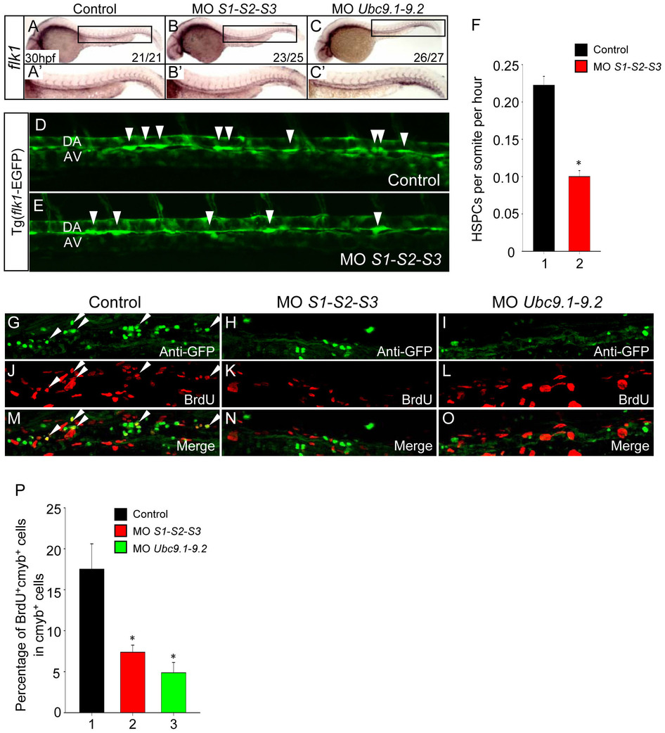
Fig. 3 Loss of SUMO reduces HSPC generation and proliferation.
(A–C) The vasculature is intact in SUMOs or Ubc9 morphants. WISH assay of flk1 at 30 hpf. (A′–C′) Magnified images of corresponding boxed regions from A to C, respectively. (D–E) The frequency of HSPC generation was reduced in SUMO-deficient embryos. Time-lapse confocal imaging analyses of HSPC generation from Tg (flk1-EGFP) line from 30 to 50 hpf. Representative pictures were captured from Video S1 and Video S2, respectively. Arrowheads indicate the budding cells. DA, dorsal aorta; AV, axial vein. (F) Statistical analyses of the frequency of HSPC generation. Data shown are the mean ± SEM, n = 3, *P < 0.01 by student′s t-test. (G–O) The proliferation of HSPC is reduced in SUMOs or Ubc9 morphants. Double immunostaining of cmyb-EGFP (G–I) and BrdU (J–L) in the CHT of Tg (cmyb-EGFP) line at 72 hpf. The bottom panel shows merged images (M–O). (P) Quantification of BrdU and cmyb-EGFP double positive cells in the CHT of Tg (cmyb-EGFP) line at 72 hpf. Data shown are the mean ± SD, n e 3, *P < 0.01 by student′s t-test.